
ملف:Brain trauma CT.jpg
لا توجد دقة أعلى متوفرة.
Brain_trauma_CT.jpg (525 × 572 بكسل حجم الملف: 59 كيلوبايت، نوع MIME: image/jpeg)
| هذا ملف من ويكيميديا كومنز. معلومات من صفحة وصفه مبينة في الأسفل. كومنز مستودع ملفات ميديا ذو رخصة حرة. |
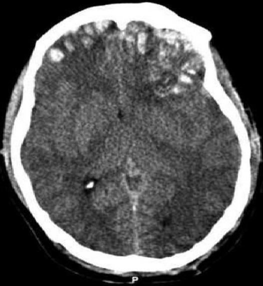
| الوصف |
Čeština: Kontuze na CT snímku se subdurálním krvácením a frakturami.
English: CT scan of patient with brain trauma. Caption reads, "Preoperative CT scan of patient while he had a GCS of 14." Accompanying text in article reads, "Emergent CT imaging revealed a sagittally oriented skull fracture extending from the vertex to the foramen magnum as well as a transverse parietal and temporal bone fracture. Multiple frontal, parietal, and temporal lobe contusions with associated interhemispheric hemorrhage and a left-sided subdural hematoma measuring 1.7 mm in greatest depth were appreciated. Effacement of the basilar cisterns was noted without shift of midline structures." |
| التاريخ | Published: 2 October 2008 |
| المصدر | Rehman T, Ali R, Tawil I, Yonas H (2008). "Rapid progression of traumatic bifrontal contusions to transtentorial herniation: A case report". Cases journal 1 (1): 203. doi:10.1186/1757-1626-1-203. PMID 18831756. http://www.casesjournal.com/content/1/1/203 PMC 2566562 |
| المؤلف | Rehman T, Ali R, Tawil I, Yonas H |
| الترخيص (إعادة استخدام هذا الملف) |
هذا الملف مُرخَّص برخصة المشاع الإبداعي العامة المُلزِمة بنسب العمل إلى مُؤَلِّفه 2.0
|
تاريخ الملف
اضغط على زمن/تاريخ لرؤية الملف كما بدا في هذا الزمن.
| زمن/تاريخ | صورة مصغرة | الأبعاد | مستخدم | تعليق | |
|---|---|---|---|---|---|
| حالي | 10:52، 3 فبراير 2023 | | 525 × 572 (59 كيلوبايت) | Doc James | Cropped 18 % horizontally, 8 % vertically using CropTool with precise mode. |
| 03:31، 24 أكتوبر 2008 |  | 640 × 619 (47 كيلوبايت) | Delldot | {{Information |Description=CT scan of patient with brain trauma. Caption reads, "Preoperative CT scan of patient while he had a GCS of 14." Accompanying text in article reads, "Emergent CT imaging revealed a sagittally oriented skull fracture extending |
استخدام الملف
ال3 صفحات التالية تستخدم هذا الملف:
الاستخدام العالمي للملف
الويكيات الأخرى التالية تستخدم هذا الملف:
- الاستخدام في az.wikipedia.org
- الاستخدام في be-tarask.wikipedia.org
- الاستخدام في be.wikipedia.org
- الاستخدام في bn.wikipedia.org
- الاستخدام في ca.wikipedia.org
- الاستخدام في cs.wikipedia.org
- الاستخدام في da.wikipedia.org
- الاستخدام في el.wikipedia.org
- الاستخدام في en.wikipedia.org
- الاستخدام في es.wikipedia.org
- الاستخدام في eu.wikipedia.org
- الاستخدام في fa.wikipedia.org
- الاستخدام في fi.wikipedia.org
- الاستخدام في fr.wikipedia.org
- الاستخدام في he.wikipedia.org
- الاستخدام في hy.wikipedia.org
- الاستخدام في id.wikipedia.org
- الاستخدام في it.wikipedia.org
- الاستخدام في ja.wikipedia.org
- الاستخدام في ko.wikipedia.org
- الاستخدام في ky.wikipedia.org
- الاستخدام في nl.wikipedia.org
- الاستخدام في pl.wikipedia.org
- الاستخدام في pt.wikipedia.org
- الاستخدام في ru.wikipedia.org
- الاستخدام في sr.wikipedia.org
- الاستخدام في ta.wikipedia.org
- الاستخدام في tr.wikipedia.org
- الاستخدام في uk.wikipedia.org
- الاستخدام في uz.wikipedia.org
- الاستخدام في www.wikidata.org
- الاستخدام في zh.wikipedia.org