
قائمة الأمراض والاضطرابات الجلدية

تصيب الكثير من الأمراض الجهاز اللحافي في الإنسان — نظام الأعضاء الذي يغطي كامل الجسم ويتكون من الجلد والشعر والأظافر وكذلك والعضلات والغدد المرتبطة به.[1] الوظيفة الرئيسية لهذا الجهاز هو أن يكون حاجزا للبيئة الخارجية.[2] معدل وزن الجلد يبلغ 4 كيلوغرامات تقريبا ويغطي مساحة مترين مربعين ويتكون من 3 طبقات متميزة هي: البشرة والأدمة ونسيج تحت الجلد.[1]
هنالك نوعان من الجلد عند الإنسان هما: الجلد المشعر والجلد الأجرد، وهو الجلد الفاقد للشعر في راحة اليد وأخمص القدم (ويسمى كذلك بالسطح «الراحي الأخمصي»).[3] في الجلد المشعر، يوجد الشعر في تراكيب تسمى بالوحدات الشعرية الزهمية، كل واحدة تحتوي على بصيلة الشعرة وغدة زهمية وعضلة ناصبة للشعر مرتبطة بها.[4] تتكون البشرة والشعر والغدد في مرحلة الجنين من الأديم الظاهر، والذي يتأثر كيميائيا الأديم المتوسط والواقع تحته والذي يكون الأدمة والأنسجة تحت الجلد.[5][6][7]
البشرة هي الطبقة الخارجية في الجلد، وهي ظهارة حرشفية مكونة من بعض الطبقات: الطبقة المتقرنة والطبقة الصافية والطبقة الحبيبية والطبقة الشائكة والطبقة القاعدية.[8] تصل التغذية إلى هذه الطبقات من الأدمة عن طريق الانتشار لكون البشرة خالية من الأوعية الدموية. تحتوي البشرة على 4 أنواع من الخلايا: الخلايا الكيراتينية والخلايا الميلانينية وخلايا لانغرهانس وخلايا ميركل. تعد الخلايا الكيراتينينة المكون الرئيسي من بين هذه الأنواع، حيث تشكل تقريبا 95٪ من البشرة.[3] يحافظ على هذه الظهارة الحرشفية المطبقة من خلال انقسام الخلايا في الطبقة القاعدية، حيث أن الخلايا المتمايزة تزاح تدريجيا من الطبقة الشائكة نحو الطبقة المتقرنة، حيث يتم التخلص من الخلايا من هذه الطبقة باستمرار.[3] معدل إنتاج الخلايا في الجلد الطبيعي يساوي معدل الفقدان، وتحتاج الخلية أسبوعين للانتقال من طبقة الخلايا القاعدية إلى طبقة الخلايا الحبيبية، وتحتاج إسبوعين إضافيين لكي تعبر الطبقة المتقرنة.[9]
الأدمة هي طبقة في الجلد تقع بين البشرة ونسيج تحت الجلد، وتتكون من مقطعين هما الأدمة الحليمية والأدمة الشبكية.[10] تتطابق الأدمة الحليمية السطحية مع حروف شبكة الأدمة التي تعلوها، وتوجد منطقة الغشاء القاعدي بين الطبقتين وتتفاعلان من خلالها.[10] تتكون الأدمة من تراكيب هي الكولاجين وألياف مرنة ومادة أساسية.[10] توجد داخل هذه المكونات الوحدات الشعرية الزهمية والعضلات الناصبة والغدد المفرزة والغدد المفترزة.[8] تحتوي الأدمة على شبكتين وعائتين تجريان بشكل متواز إلى سطح الجلد، إحداهما واحدة سطحية والأخرى ضفيرة عميقة، وهما مرتبطتان بأوعية موصلة عمودية.[8][11] وظيفة الأوعية الدموية في الدمة مكونة من أربعة فقرات هي: توفير التغذية وتظيم درجة الحرارة وتنظيم الاتهاب والمشاركة في شفاء الجروح.[12][13]
نسيج تحت الجلد هو طبقة دهنية تقع بين الأدمة واللفافة الواقعة تحتها.[14] هذا النسيج بدوره ينقسم إلى مكونين هما: الطلبقة الدهنية الحقيقة أو السبلة الشحمية، وتوجد تحتها طبقة أثرية من العضلات هي السبلة العضلية.[3] المكونات الخلوية الرئيسية في هذا النسيج هي الخلايا الدهنية.[14] يتكون تركيب هذا النسيج من حيز حاجزي وحيز فصي، وهما يختلان في المظهر تحت المجهر.[8] تتضمن وظيفة الطبقة الدهنية تحت الجلد عزل الجسم وامتصاص الإصابات وتوفر الحفظ لمصادر الطاقة.[14]
الحالات المرتبطة بالجهاز اللحافي عند الإنسان تتضمن طيفا واسعا من الأمراض، يطلق عليها تسمية الجلادات، بالإضافة للكثير من الحلات غير المرضية مثل (كما في ظروف معينة: فرط ميلانين الظفر وأظافر المضرب).[15][16] ورغم أن هنالك عددا قليلا من الأمراض مسجلة لأغلب زيارات المرضي لأطباء الجلد، لكن هنالك آلاف الحالات الجلدية تم العرف عليها ووصفها.[14] ويواجه تصنيف هذه الأمراض تحديات في علم تصنيف الأمراض، حيث أن الكثير من أسباب هذه الأمراض ومرضيتها عادة غير معروفة.[17][18] لذلك فإن أكثر الكتب العلمية تصنف هذه الأمراض حسب موقعها وتكوينها وأسبابها وغيرها.[19][20] يجرى التشخيص السريري لأي حالة جلدية معينة من خلال جمع معلومات المريض المتعلقة بالآفة الجلدية وتشمل موقعها (مثل الذراع والرأس والساق) والأعراض (الحكة والألم) والمدة (حادة أو مزمنة) والتنسيق (منعزل أو عام أو حلقي أو خطي)، والتشكل (البقع والحطاطات والحويصلات) واللون (أحمر أو أزرق أو بني أو أسود أو أبيض أو أصفر).[21] يحتاج تشخيص الكثير من الأمراض إلى خزعة جلدية لتعطي المعلومات النسيجية[22][23] التي يمكن ربطها مع التشخيص السريري والنتائج المختبرية.[24][25][26]
طفح عدي الشكل (أو على شكل حبوب)[عدل]
يعود ظهور الطفح عدي الشكل إلى التغيرات في الوحدة الشعرية الزهرية.[27][28]
- حب الشباب الصيفي[nb 1][nb 2][nb 3]
- حب الشباب المكبب
- حب شباب مستحضرات التجميل
- حب الشباب المداهم
- حب الشباب الجادر العنقي القفوي

- حب شباب الميكانيكي
- حب الشباب الدوائي
- حب الشباب الجاروسي النخري
- حب الشباب الشائع
- حب الشباب مع وذمة الوجه[nb 4]
- تعجر الجفن
- العد الوردي متوسع الشعيرات الحمراء
- حب الشباب العصبي السحجي (حب الشباب شجب الفتيات أو حب شباب بيكر)[nb 5]
- العد الوردي الغدي
- تعجر الفك
- عد وردي سلبي الغرام
- التهاب الجلد الوجهي الحبيبي

- التهاب الجلد حول الفم الحبيبي
- حب الشباب الهالوجيني
- التهاب الغدد العرقية القيحي
- الورم الحبيبي العقيم الوجهي مجهول السبب
- حب الشباب الطفيلي
- العد الوردي الذنبي الشكل
- الذئبة الدخنية الوجهية المنتشرة
- تعجر الجبهة
- حب الشباب الوليدي
- حب الشباب المهني
- حب الشباب النفطي
- العد الوردي العيني
- تعجر الأذن
- التهاب جلد حول الفوهة
- وذمة العد الوردي المستديمة
- عد وردي تعجري
- عد المراهم (أو حب شباب المراهم)
- عد وردي حطاطي بثري (أو عد وردي التهابي)
- التهاب حوائط الجريبيات الرأسية المتقح الخانق أو التهاب هلل الفروة السالخ
- التهاب الجلد حول الفم
- التهاب محيط الحجاج
- تقيح جلد الوجه (أو العد الخاطف)
- تعجر الأنف
- عد وردي
- عد وردي مكبب
- متلازمة سافو (مشتقة من متلازمة التهاب الزليل–حب الشباب–البثار–فرط التعظم–التهاب العظام)[nb 6]
- عد وردي ستيرويدي
- حب الشباب القطراني
- حب الشباب المداري
متلازمات التهابية ذاتية[عدل]
المتلازمات الالتهابية الذاتية هي مجموعة من الاضطرابات الوراثية التي تتميز بنوبات آفات جلدية التهابية وحمى دورية.[29][30]
- متلازمة بلاو
متلازمة بلاو - المتلازمة الجلدية والمفصلية العصبية الطفولية المزمنة (أو الداء الالتهابي متعدد الأجهزة وليدي البدء)
- شرى البرد العائلي
- حمى البحر الأبيض المتوسط
- فرط الغلوبولين المناعي د في الدم[nb 7]
- متلازمة مجيد
- متلازمة ماكل - ويلز
- متلازمة الحمى الدورية المرتبطة بمستقبل عامل نخر الورم (أو الحمى الهيبرنية العائلية)

تبثر مزمن[عدل]
حالات ظهور التقرحات الجلدية المزمنة والتي تستمر لفترة طويلة مع وجود حويصلات وفقاعات.[31][32][33]
- داء الغلوبولين المناعي أ الخطي للبالغين
- الفقعان الفقاعي
- داء الغلوبولين المناعي أ الخطي للأطفال (الداء الفقاعي المزمن عند الأطفال)
- شبيه الفقاع الندبي
- التهاب الجلد الحلئي الشكل
- الفقاع المتوطن
- انحلال الجلد الفقاعي المكتسب
- داء غروفر (أو الجلاد الحال للأشواك العابر)
داء غروفر - فقاع الغلوبولين المناعي أ
- جلاد الغلوبولين المناعي أ بالعدلات داخل البشرة
- شبيه الفقاع الندبي الموضعي
- فقاع الأباعد الورمية
- فقاع حمامي (متلازمة سينيار أوشر)
- الفقاع القرطاسي
- فقاع هربسي الشكل
- فقاع عقيدي
- فقاع تنبتي
- فقاع تنبتي نمط هالوبو
- فقاع تنبتي نمط نيومان
- الفقاع الشائع
- فقاع حويصلي
- شبيه فقاع الطفولة الفرجي

أمراض الأغشية المخاطية[عدل]
حالات الأغشية المخاطية تتضمن الأغشية المبطنة الرطبة للعين والأنف والفم والأعضاء التناسلية والشرج.[34]

- انعدام الكاتالاز (داء تاكاهارا)
- صداف مختل التقرن المكتسب
- التهاب الشفة السفعي
- التهاب اللثة التقرحي الناخر الحاد
- التهاب الشفة التماسي الأرجي
- الذبحة الفقاعية النزفية
- التهاب الشفة الزاوي
- مرض بهجت
- اللسان الأسود المشعر
- لسان كافيار
- التهاب الشفة التقشري
- التهاب الشفة الغدي
- التهاب الشفة الورمي الحبيبي
- الجيوب السنية
- قلة العدلات الدورية
- التهاب اللثة التوسفي
- قرحة الشفة المحدثة بالأدوية
- تشكل بشرة الشفة
- ورم لثوي
- الورم اللثوي التشققي
- التهاب الحليمات اللسانية الطفحي
- تنسج أحمر
- اللسان المشقق
- اللسان الجغرافي
- ورم ليفي لثوي
- تضخم اللثة
- طلاوة مشعرة
- جيب سني داخل الفم
- الخط الأبيض
- الصداف أو الطلوان
- صداف مع ثفان وسرطانة مريئية
- قرحة فموية كبرى (أو التهاب حوائط الغدة المخاطي النخري الناكس)
- التهاب اللسان المعيني الناصف
- آفة فموية ميلانينية
- متلازمة ميلكرسون روزنتال
- مضغ الخد المزمن (أو عض الخد المزمن)
- سرطانة حرشفية الخلايا المخاطية
- قيلة مخاطية فموية (أو سليلة مخاطية فموية أو الكيس المخاطي للغشاء المخاطي للفم)
- بقع ناغاياما
- داء كرون الفموي
- الورام الحليمي المزهر الفموي
- فرط الميلانين الفموي
- الورم الأرومي الاغترابي العظمي اللساني
- الورم الأرومي المينائي المحيطي
- التهاب الشفة بالخلية البلازمية (أو التهاب اللثة بالخلية البلازمية)
- ورم شائكي بلازمي
- طلوان ثؤلولي تكاثري
- ورم حبيبي مقيح

- التهاب الفم القيحي التنبتي
- التهاب الفم القلاعي الراجع
- عدوى الهربس البسيط داخل الفم الراجعة
- اللسان الناعم (أو التهاب اللسان الضموري)
- التهاب الفم النيكوتيني
- نتوء حنكي
- ثؤلول البواق
- ورم حليمي دهليزي
- وحمة إسفنجية بيضاء
أمراض لواحق الجلد[عدل]
إن حالات لواحق الجلد هي الحالات التي تؤثر على غدد الجلد والشعر والأظافر والعضلات الناصبة للشعر.[1][35]
- حب الشباب النخري أو العد النخري
- فرط الشعر المتعمم المكتسب (أو فرط الشعر الزغابي المكتسب)
- الجلاد الثاقب المكتسب (أو الداء الكولاجيني الثاقب المكتسب)
- تقران نهايات الأباعد الورمية (أو متلازمة بازكس)
- انحلال عظام النهايات
- التقرح حول الظفري

- ثعلبة مبقعة
- ثعلبة مسرطنة (أو ثعلبة مورمة)
- تشاقط الشعر المتنامي
- ثعلبة ذكرية الشكل
- نقص التعرق (أو اللاعرقية)
- انعدام الأظفار
- الوبش الظاهري
- خطوط بو
- الأظفار الزرقاء (أو الهليلات الزرقاء أو الازوردية)
- الصنان (عرق الإبط أو نتن العرق أو رائحة التعرق)
- تشوه الشعر الفقاعي
- ثعلبة ندبية نابذة مركزية (أو ثعلبة المشط الساخن أو متلازمة التنكس الجريبي أو الثعلبة الكاذبة لمركز فروة الرأس)
- ظفر شيفرون
- تعرق ملون
- التقرح حول الظفري المزمن
- ثعلبة ندبية
- تعجر الأصابع
- زاوية لوفيبوند
- خلل تنسج السبابة الخلقي
- التهاب الجريبات القمعية المتكرر والمنتشر
- جلاد فروة الرأس البثري المؤتكل (أو التهاب جلد فروة الرأس البثري المؤتكل)
- فرط الميلانين الأحمر الجريبي في الوجه والرقبة
- التهاب الجريبات الصالع
- التهاب الجريبات المنخر الثاقب
- داء فوكس-فوردايس
- الثعلبة المليفة الأمامية (أو الجبهية)
- فرط الشعر المتعمم الخلقي
- فرط التعرق المتعمم
- متلازمة غراهام ليتل
- حباب أنفي أحمر

- الأظفار الخضراء
- فرط التعرق تذوقي المنشأ
- صؤاب كاذب (أو مصبوب الشعر)
- وحمة مسام الشعر (ورم الزغب العابي)
- باطن الراحة والأخمص المشعرة
- أظفار ليندسي
- ظفر معلق
- تلين الأظفار
- تعرق مدمي
- شعرانية
- ظفر شصي
- ثعلبة المشط الساخن
- فرط أشعار المرفق (متلازمة المرفق المشعر)
- فرط أشعار فروة الرأس البسيط
- حثل حويصلة الشعر المتقطع
- تقران الجريبات الشعرية المضمر
- شعر متلوي (تلوي الشعر المترقي المكتسب)
- ورم كونن (أو ورم ليفي تحت الظفر)
- تقعر الأظفار
- داء كيرل (أو فرط التقران الجريبي والمجاور للجريبات الثاقب للجلد)
- الوبش (أو الأظفار البيضاء)
- حزاز مسطح جريبي
- حزاز مسطح في الأظفار
- حزاز شائك (أو تقران شوكي)
- ثعلبة متشحمة (أو فروة الرأس المتشحمة)
- فرط الشعر المكتسب الموضعي
- فرط الشعر الخلقي الموضعي
- احمرار الأظفار الطولاني
- فرط ميلانين الظفر الطولاني
- متلازمة طور التنامي الرخو (أو متلازمة طور تنامي الشعر الرخو)
- الذئبة الحمامية
- مرط الجفنين
- سوء ترصيف صفيحة الظفر
- قرعة ذكرية النمط
- نقص شعر ماري أونا (أو نقص شعر ماري أونا الوراثي)
- حثل الظفر الأوسط (أو حثل الظفر المخدود الناصف أو حثل الظفر المخدود الناصق لهلر أو الظفر المخدود أو حثل الظفر الأخدودي)
- خطوط ميس
- فرط ميلانين الظفر
- داء منكس (أو متلازمة منكس أو داء الشعر المتلوي أو داء الشعر المفتل أو متلازمة منكس للشعر المتلوي أو المفتل)
- تعجر الشعر (الشعر المعجر)
- وبش مخطط (أو أظفار موركه أو خطوط موركه)

- متلازمة الظفر والرضفة (أو متلازمة فونغ أو خلل تنسج المفاصل والأظفار العظمي الوراثي أو متلازمة هود)
- أورام فراش الظفر
- فرط الشعر الوحماني
- ثعلبة لاندبية
- ضخامة الظفر
- ضمور الظفر
- نشوب الظفر (أو الظفر الناشب)
- انعقاف الأظفار
- انفكاك الظفر
- تساقط الأظفار
- ورم مطرس الظفر
- قضم الظفر (أو قضم الأظفار)
- تقرن فراش الظفر
- تمرط الأظفار (أو ثعلبة الأظفار)
- تكسر الأظفار (أو الأظفار الهشة)
- تشقق الأظفار
- هوس نتش الأظفار
- ثعلبة بقعية
- فرط التعرق الراحي الأخمصي (أو فرط التعرق الانفعالي)
- نظير التقرن البثري
- فرط الشعر المكتسب المنطبع
- التهاب الجريبات الثاقب
- شعر محلقن
- انشعاب الشعر
- أشعار توائم
- شعر محلقن كاذب
- شعر ملتوٍ
- أظفار الملقط (أو أظفار أوميغا أو أظفار ترمبت)
- نخالية أسبستية الشكل (أو سعفة أسبستية الشكل)
- تقوس الأظفار
- ثنية اعتلال عصبي (أو شعر متلبد)
- ظفر بلومر
- فرط الشعر سابق البلوغ
- ثعلبة الضغط (أو ثعلبة تالية للجراحة أو ثعلبة محدثة بالضغط)
- التهاب جريبات شعر اللحية الكاذب (أو حكة الحلاق أو التهاب جريبات شعر اللحية الرضحي أو كدمات الحلاقة أو التهاب جريبات شعر اللحية الكاذب التندبي)
- ثعلبة بروك الكاذبة (أو ثعلة ندبية)
- أظفار الصدفية
- ظفرة منقلبة ظفرية (أو ظفرة بطنية)
- ظفرة ظفرية (أو ظفرة ظهرية)
- فرفرية فراش الظفر
- ظفر المضرب (أو قصر الأظفار)
- التهاب الغدد العرقية الراحية الأخمصية المتكرر (أو التهاب الغدد العرقية الراحية الأخمصية مجهول السبب أو التهاب الغدد العرقية الأخمصية مجهول السبب أو حمامى أخمصية مؤلمة أو التهاب الغدد العرقية الراحية الأخمصية الناتح أو التهاب السبلة الشحمية الأخمصية)
- هليلات حمراء
- متلازمة روس
- متلازمة روبينشتاين - تايبي
- متلازمة ستليس
- متلازمة الظفر الصدفة
- متلازمة طور التنامي القصير
- نزف شظوي

- هليلات مبقعة
- تلون صفيحة الظفر
- أظفار مرقطة
- الورم الدموي تحت الظفر
- تساقط الشعر الكربي
- أظفار تيري
- ثعلبة الشد
- ثعلبة رضحية
- داء الجريبات الوزي الرضحي
- ثعلبة مثلثة (أو ثعلبة صدغية أو ثعلبة مثلثة صدغية)
- ضخامة شعرية
- داء الفطريات الشعرية الإبطي
- تقصف الشعر الانغلافي (أو شعر خيزراني)
- تقصف الشعر العقد
- الركود الشعري الشوكي
- التهاب الجريبات الملفوفة
- ثعلبة ورمية
- حثل الأظفار العشرين (أو الأظفار المصقولة أو الظفر المخدد)
- متلازمة الشعر غير القابل للتمشيط
- وحمة الشعر الصوفي
- فرط الشعر المرتبط بالكروموسوم أكس
أمراض طبقة تحت الجلد الدهنية[عدل]
حالات دهون تحت الجلد هي الحالات التي تصيب طبقة النسيج الدهني الواقع بين الأدمة واللفافة الواقعة تحتها.[36][37][38][39]
- الحثل الشحمي المكتسب المتعمم (أو متلازمة لورنس أو متلازمة لورنس-سيب)
- الشحامة المؤلمة (أو داء دركوم)
- التهاب السبلة الشحمية بنقص ألفا 1-أنتيتريبسين
- التهاب السبلة الشحمية بضمور النسيج الضام
- متلازمة باراكيه-سيمونز (الحثل الشحمي الجزئي المكتسب أو الحثل الشحمي الرأسي الصدري أو الحثل الشحمي المترقي)
- ورام شحمي متناظر حميد (أو داء مادلونغ)
- الحثل الشحمي البطني النابذ (أو الحثل الشحمي النابذ أو الحثل الشحمي النابذ البطني الطفولي)
- حمامى عقدة مزمنة (أو حمامى عقدة هاجرة أو التهاب السبلة الشحمية العقيدي الجوال تحت الحاد لفيلانوفا وبينيول أو التهاب السبلة الشحمية العقيدي الجوال تحت الحاد)

- التهاب السبلة الشحمية البارد
- الحثل الشحمي الخلقي المتعمم (أو متلازمة بيراردينلي-سيب)
- التهاب السبلة الشحمية المنسج الملتهم للخلايا
- حثل شحمي محدث بالدواء
- التهاب السبلة الشحمية المفتعل
- الحثل الشحمي العائلي الجزئي (متلازمة كوبرلنغ-دونيغان)
- التهاب السبلة الشحمية النقرسي
- متلازمة فرط التنسج الشقي والورام الشحمي المتعدد
- الحثل الشحمي المرتبط بفيروس العوز المناعي البشري[nb 8]
- ضمور شحمي أوبي
- الضمور الشحمي الحلقي (ضمور فيريرا-ماركيز الشحمي)
- الضمور الشحمي الهلالي
- التصلب الجلدي الشحمي (أو التهاب السبلة الشحمية المزمن مع تغيرات غشائية شحمية أو التهاب السبلة الشحمية المتصلب أو ركود التهاب السبلة الشحمية)
- تضخم شحمي
- الحثل الشحمي الموضعي
- التهاب السبلة الشحمية الفصي بالعدلات
- التهاب وعائي عقدي
- ضمور شحمي نصف وجهي خطي متأخر الظهور غير مترقي
- التهاب السبلة الشحمية البنكرياسي (أو التهاب السبلة الشحمية الإنزيمي أو تنخر الدهن البنكرياسي أو تنخر دهن تحت الجلد)
- متلازمة بولاند
- التهاب السبلة الشحمية بعد الستيرويد
- صلدمة الولدان
- ورم حبيبي شحمي متصلب (ورم برافيني)
- التهاب السبلة الشحمية الحاجزي
- تنخر دهن تحت الجلد لحديثي الولادة
- التهاب السبلة الشحمية الرضحي
- متلازمة انحلال الورم
- داء ويبر-كريستيان (التهاب السبلة الشحمية اللاقيحي الحموي المنتكس)
العيوب الخلقية[عدل]
العيوب الخلقية الجلدية هي مجموعة متنوعة من الاضطرابات الناتجة عن عيوب في التشكل الحيوي، وهي العملية الحيوية في تكوين شكل جسم الإنسان.[35][40][41]
- ظفر إضافي في أصبع القدم الصغير
- زنمة زائدة (أو زائدة أمام الأذن)
- متلازمة الشريط السلوي
- متلازمة الشريط السلوي
- التوصيلة الشريانية الوريدية
- ورم وعائي وليدي حميد
- كيسة الفلح الخيشومي
- كيسة قصبية المنشأ
- ورم وعائي شعيري
- تشوه وريدي كهفي
- راحة العنق الغضروفية العنقية (زنمة عنقية زائدة أو غبب أو لغد)
- جلاد حويصلي ومؤتكل خلقي
- تضخم طية الإبهام الوحشية الخلقي
- حفرة الشفة الخلقية
- تشوه تقاطع النهايات الخلقي
- ورم العضلة الملساء العابي الخلقي
- تشوه لمفي كيسي
- كيسة جلدانية
- ورام وعائي وليدي منتشر
- قيلة دماغية
- خلل تنسج جلدي وجهي بؤري
- أسنان هتشنسون
- تشوه شعيري وريدي جلدي مفرط التقرن
- نخر بشروي داخل الرحم
- المتلازمة الطرفية الثديية
- متلازمة لوري ماكلين
- ضخامة الشفاه
- تشوه لمفي كيسي كبير (ورم رطب كيسي)
- ورم المطرس الشعري الكبير (أو سرطانة المطرس الشعري)
- الداء الفقاعي بالمناعة الذاتية الأمومي
- كيس رفاء وسطي
- ورم الأديم العصبي الظاهر الميلانيني عند الرضع
- عدم تنسج الجلد الغشائي
- تشوه لمفي تكسي دقيق
- فلح عنقي ناصف
- بقعة منغولية (كثرة الخلايا الملانينية الجلدية الخلقي أو كثرة الخلايا الملانينية الجلدية)

- رحى توتية (ج. أرحاء توتية)
- خلل التعظم القمي والجهي الناجري
- ورم دبقي أنفي (انتباذ شبيه بالدماغ أو انتباذ رأسي شبيه بالدماغ أو ورم عابي دبقي أو نسيج دبقي عصبي نابذ أو انتباذ دماغي أنفي أو نسيج دماغي نابذ أنفي)
- كيسة القناة الأنفية الدمعية
- وحمة ذربية دهنية
- ورم وعائي خلقي غير ملتف
- كيسة القناة السرية المساريقية (أو بقية القناة السرية المساريقية أو كيسة محية)
- متلازمة بيلفيس
- ورم المطرس الشعري (أو الورم الظهاري المكلس لمالرب)
- داء بولاند
- متلازمة فاسيس (أو متلازمة تشوه الحفرة الخلفية وأورام وعائية وشذوذ الشرايين وعيوب قلبية وتشوهات العين وفلح قصي ورفاء فوق السرة)
- كيس وجيب أمام الأذن (وهدة الأذن أو ناسور خلقي أذني أو ناسور خلقي أمام الأذن أو كيس أمام الأذن)
- ورم وعائي خلقي سريع الالتفاف (ورم وعائي غير مترقي خلقي)
- متلازمة روزنتال-كلوبفر
- أصبع زائد رديم (كثرة الأصابع الرديمية)
- متلازمة ساكرال
- كيس سمحاق القحف
- رصعة جلدية (حفرة جلدية)
- تشوه لمفي سطحي (ورم وعائي لمفي متحدد)
- حلمة زائدة (أو حلمة كاذبة)
- كيس درقي لساني
- تشوه وعائي ثؤلولي (تقران وعائي متحدد وحمي الشكل)
أمراض النسيج الضام[عدل]
تعود أسباب أمراض وحالات النسيج الضام لمجموعة معقدة من الاستجابات مناعية ذاتية التي تستهدف أو تؤثر على الكولاجين أو المادة القاعدية.[35][42]
- الذئبة الحمامية الجلدية الحادة (أو تصلب الجلد شاذ الضباغ والضموري أو قشيعة مسطحة ضمورية أو تصلب الجلد الضموري الفوري)
- متلازمة كرست (وهي مختصر لمتلازمة الكلاس–ظاهرة رينو–خلل حركة المرئ–تصلب الأصابع–توسع الشعيرات)
- الذئبة الحمامية التثليجية (أو الذئبة الحمامية التثليجية لهوتشينسون)
- التهاب العضلات والجلد اليفعي
- الذئبة الحمامية القرصية الطفولية
- الذئبة الحمامية الشاملة الطفولية
- متلازمات عوز المتممة
- التهاب العضلات والجلد
- متلازمة كثرة اليوزينيات والألم العضلي
- تصلب الجلد الخطي الجبهي
- الذئبة الحمامية القرصية المتعممة
- قشيعة متعممة
- التهاب الجلد الورمي الحبيبي الخلالي
- الالتهاب المفصلي الروماتويدي اليفعي (الالتهاب المفصلي مجهول السبب اليفعي أو داء ستِل)
- قشيعة جدروية (أو جدرة أديسون)
- ضمور الجلد الخطي لمولين
- تصلب الجلد الخطي
- الذئبة الحمامية القرصية الموضعية
- قشيعة موضعية
- التهاب السبلة الشحمية بالذئبة الحمامية (الذئبة الحمامية العميقة أو التهاب السبلة الشحمية الذئبي أو الذئبة العميقة أو الذئبة الحمامية الجلدية)
- متلازمة الذئبة الحمامية وتراكب الحزاز المسطح
- طفح بقعي حطاطي محدث بميثوتركسيت
- مرض النسيج الضام المختلط (أو متلازمة شارب أو مرض النسيج الضام غير المتمايز)
- قشيعة عميقة
- تراكب حزاز متصلب وضموري وقشيعة
- متلازمة القرحة الفموية والتناسلية مع الغضروف الملتهب (أو متلازمة ماجيك)
- الذئبة الحمامية الوليدية
- تليف جهازي كلوي المنشأ (اعتلال الجلد المتليف كلوي المنشأ)
- متلازمة نيكولاس-بالوس

- متلازمة التهاب العقيدات والاعتلال المفصلي وانحلال العظم
- الكلاس الورمي العائلي سوي فوسفات الدم
- التهاب الجلد بالعدلات والورمي الحبيبي السياجي
- قشيعة متصلبة شاملة
- متلازمة باري رومبرج (الضمور النصف وجهي التصاعدي)
- التصلب المجموعي المترقي
- التهاب الغضاريف الناكس (أو التهاب الغضاريف الضموري أو التلين الغضروفي المجموعي)
- التهاب المفاصل الروماتويدي
- التهاب العقيدات الروماتويدي (أو التهاب العقيدات الروماتويدي المعجل)
- التهاب وعائي الروماتويدي
- متلازمة رويل
- الوذمة الصلبة في البالغين (أو داء بوشكه أو وذمة صلبة سكرية المنشأ أو الوذمة الصلبة في البالغين لبوشكه أو الوذمة الصلبة لبوشكه)
- سحار سيليسي
- متلازمة شوغرن (أو داء ميكوليتس أو متلازمة سيكا)
- الذئبة الحمامية الجلدية تحت الحادة
- الذئبة الحمامية الشاملة
- متلازمة الزيت السام
- الذئبة الحمامية الورمية
- متلازمة توزون (أو ضمور الجلد دودي الشكل)
- الذئبة الحمامية الثؤلولية (أو الذئبة الحمامية الضخامية)
- متلازمة وينتشستر
شذوذ الأنسجة الليفية والمرنة الجلدية[عدل]
يعود سبب شذوذ الأنسجة الليفية والمرنة الجلدية لمشاكل في تنظيم تخليق الكولاجين أو تحطيمه.[35][43]

- التهاب جلد الأطراف الضموري المزمن (أو داء هركسهايمر أو الضمور المنتشر الأولي)
- تنكس النسيج المرن السفعي (أو تنكس النسيج المرن الشمسي)
- ضمور الجلد البقعي
- ارتخاء الجفن
- تهدل الجلد (أو ارتخاء الجلد أو انحلال النسيج المرن المتعمم أو تمزق النسيج المرن المتعمم أو القيلة الجلدية الثخينة)
- جلد القفا المعيني التقسيمات
- متلازمة إهلرز-دانلوس (أو جلد مفرط المرونة أو الجلد المطاطي أو الجلد المطاطي الهندي)
- تنكس النسيج المرن الساعي الثاقب
- بيلة هوموسيستينية
- ضمور الجلد البقعي لياداسون-بيليتساري
- تنكس النسيج المرن البؤري الخطي (أو الخطوط المرنة)
- متلازمة لويز- ديتز
- متلازمة مارفان
- متلازمة القرن القذالي
- تكون العظم الناقص (أو متلازمة لوبشتاين)
- تنكس النسيج المرن الثاقب الكلسي (أو الورم الأصفر الكاذب المرن الجلدي المكتسب الموضعي أو تنكس النسيج المرن الكلسي الثاقب المحيط بالسرة)
- صفروم كاذب مرن (أو الورم الأصفر الكاذب المرن أو متلازمة غرينبلاد-ستراند)
- الداء الكولاجيني الثاقب التفاعلي
- ضمور الجلد البقعي لشفيننغر-بوزي
- الورم الليفي التصلبي
- الخطوط المضمرة
- الخطوط التوسعية
- داء أولريش
- ورم كولاجيني ثاقب ثؤلولي
- متلازمة الجلد المجعد
نمو الأدمة وتحت الجلد[عدل]
يحدث نمو الأدمة وتحت الجلد بسبب:
- تكاثر تفاعلي أو ورمي لمكونات خلوية في الأدمة أو نسيج تحت الجلد.
- ورم يستهدف الأدمة أو يوجد فيها بشكل شاذ.[1][35]
- ورم وعائي لمفي مترق مكتسب (أو ورم وعائي لمفي بطاني حميد)
- توسع نهايات الشرايين
- ورم قرني ليفي طرفي (أو ورم قرني ليفي أصبعي مكتسب أو ورم قرني ليفي محيط بالظفر مكتسب)
- ثؤلول معنق (أو زائدة جلدية أو ورم حليمي جلدي أو طغوة جلدية أو سليلة ليفية ظهارية أو، ورم ليفي مليسائي أو ورم ليفي متدل أو ورم حليمي عنقي أو ورم ليفي لين أو طغوة تمبلتون الجلدية)
- ورم غدي زهمي
- البزوغ المتعمم لكثرة الخلايا البدينة الجلدية للبالغين
- غرن أو ساركومة كابوسي الجلدية الأفريقية

- غرن أو ساركومة كابوسي بتضخم العقد اللمفية الأفريقية
- ورام ليفي طفولي عدواني
- غرن أو ساركومة كابوزي المرتبطة بالأيدز
- الأينوم (أو انحلال الأصابع التلقائي أو شوكابالا)
- ورم ليفي وعائي
- تقران وعائي
- تقران وعائي لفوردايس (تقران وعائي للصفن والفرج)
- تقران وعائي لميبيلي (ثؤلول التوسع الوعائي)
- ورم عضلي أملس وعائي
- ورم عضلي أملس وعائي شحمي
- ورم شحمي وعائي
- ورم وعائي ساعي
- ساركومة وعائية (أو غرن وعائي)
- ورم ليفي سفاقي (ورم ليفي سفاقي مكلس أو ورم ليفي سفاقي يفعي)
- ورم أصفر ليفي لانمطي أو لانموذجي
- ورام أرومي شحمي حميد (أو ورم ليفي جنيني أو مضغي)
- متلازمة بوشكه-أوليندورف (أو تليف جلدي عدسي منتشر)
- أم الدم الشعرية أو الشعيرية
- ورم سرطاوي
- ورم وعائي كرزي (أو بقعة دي مورغان أو ورم وعائي شيخوخي)
- التهاب غضروف وجلد حتار الأذن العقيدي المزمن (أو التهاب غضروف وجلد حتار الأذن العقيدي)

- ورم شحمي شبه غضروفي
- ورم حبلي
- ساركومة كابوسي معهودة
- ورم ليفي كولاجيني (أو ورم أرومي ليفي صلد)
- ورم بطاني وعائي مركب
- وحمة النسيج الضام (أو ورم كولاجيني أو ورم النسيج المرن أو لطخة حرشاء)
- انتباذ بطاني رحمي جلدي
- ورم سحائي جلدي (نسيج سحائي مغاير التوضع أو قيلة سحائية رديمية)
- تليف نقوي جلدي
- ورم مخاطي جلدي
- جلد مرمري متوسع الشعيرات خلقي (أو دوالي متعممة خلقية أو متلازمة فان لوهويزن)
- ورم عابي بالخلية المتغصنة الأدمية
- ورم ليفي جلدي (ورم المنسجات الليفي الحميد أو ورم الخلية المتغصنة الأدمية أو ورم المنسجات الليفي أو ورم المنسجات الليفي أو ورم ليفي بسيط أو ورم المنسجات أو تليف عقيدي تحت البشرة أو ورم وعائي مصلب)
- ساركومة ليفية جلدية حدبية
- ورم رباطي
- كثرة الخلايا البدينة المنتشرة في الجلد
- ورم ليفي طفولي منتشر (أو ورام ليفي طفلي منتشر)
- تفقع دوبويترن (أو أهبة دوبويترن أو داء دوبويترن أو ورام ليفي راحي)
- ورم عابي وعائي ناتح
- ورم ليفي مرن ظهري
- ورم بطاني وعائي حليمي داخلي (أو ورم دابسكا أو ورم بطاني وعائي نوع دابسكا أو ورم بطاني وعائي مسماري أو ورم بطاني وعائي حليمي داخلي خبيث أو ورم بطاني وعائي لمفي حليمي)
- ورم المنسجات للخلية الشبيهة بالظهارية
- ورم بطاني وعائي شبيه بالظهارة
- ساركومة شبيهة بالظهارة
- كثرة الخلايا البدينة المحمرة للجلد
- ورم غضروفي غير هيكلي (ورم غضروفي في الأجزاء اللينة)
- أورام ليفية وعائية مخاطية عائلية
- فتق لفافي
- ورم ليفي في غمد الوتر
- ورام ليفي رقبي (أو ورم قصي خشائي عند الرضع)
- ورم عابي ليفي عند الرضع
- حطاطة الأنف الليفية (أو الحطاطة الليفية الوحيدة الحميدة أو حطاطة الوجه الليفية)
- جلد مجعد مع تندب (متلازمة طفل عجلة ميشلان)
- داء فوردايس (أو بقعة فوردايس)
- كيسة عقدة (أو كيسة تحت الزليل)
- ورم عصبي عقدي
- ورم عضلي أملس تناسلي (أو ورم عضلي أملس سلخي)
- ورم أرومي ليفي للخلية العملاقة
- ورم الخلايا العملاقة في غمد الوتر (ورم زليلي للخلية العملاقة، أو التهاب زليل الوتر العقيدي أو التهاب زليل الوتر العقيدي المصطبغ)
- ورم وعائي شبيه كبيبي

- ورم كبي (ورم وعائي كروي أو ورم كبي صلب أو ورم كبي وحيد)
- ورم الخلية الحبيبية (ورم أبريكوسوف أو الورم الأرومي العضلي الحبيبي الخلايا أو ورم الغمد الصبي للخلايا الحبيبية أو ورم شفاني خلوي حبيبي)
- ورم عابي
- ورم الخلايا الحوطية (أو ورم الخلايا الخولية)
- ساركومة وعائية
- ورم إشتائي (أو ورم الدهن الأسمر أو ورح شحمي جنيني أو ورم دهن الجنين أو الورم الشحمي للنسيج الشحمي غير الناضج)
- ندبة ضخامية
- ساركوما كابوسي المرتبطة بكبت المناعة
- ورم ليفي أصبعي طفلي (أو ورم ليفي مشتمل أو ورم ليفي عضلي أصبعي طفلي أو ورم راي)
- ورم الخلايا الحوطية الطفلي أو الخلقي (أو ورم الخلايا الخولية الطفلي أو الخلقي)
- ورام ليفي عضلي طفلي (أو ورام ليفي متعمم خلقي أو ورام ليفي متعدد المراكز الخلقي)
- تزجج أو تنكس زجاجي جهازي طفلي (أو تزجج أو تنكس زجاجي جهازي يفعي)
- ورم الخلايا المغزلية الشحمي داخل الجلد
- فرط التنسج البطاني الحليمي الوعائي (أو آفة ماسون أو ساركومة وعائية لماسون أو ورم ماسون أو فرط التنسج البطاني الحليمي)
- ورام ليفي زجاجي (أو هياليني) يفعي (أو ورام ليفي زجاجي بسيط يفعي أو متلازمة موراي-بورتك-دريشر)
- ورم بطاني وعائي شكل كابوسي (أو ورم بطاني وعائي شكل كابوسي الوليدي)
- متلازمة كاساباك-ميريت (أو ورم وعائي مع قلة الصفيحات)
- جدرة (ندبة جدرية)

- حؤول متقرن
- كيسة قرنية
- متلازمة كيبل ترينوني ويبر (أو متلازمة كليبل-ترينوناي أو متلازمة التضحم العظمي الوعائي أو التخضم التوسع الوعائي الدموي)
- وسائد برجمية (تثفن الجلد)
- ساركومة عضلية ملساء
- ورم شحمي
- ساركومة شحمية (ورم شحمي لانمطي)
- توسع الأوعية اللمفية (أو ورم وعائي لمفي)
- ورام وعائي لمفي
- ورم المنسجات الليفي الخبيث (أو ساركومة متعددة الأشكال غير متمايزة)
- ورم الغمد العصبي المحيطي الخبيث (ورم شفاني خبيث أو ساركومة ليفية عصبية أو ساركومة عصبية)
- ساركومة الخلية البدينة
- قيلة سحائية
- سرطانة نقيلية
- ورم وعائي وريدي دقيق (ورم وعائي شعري دقيق)
- وحمة خمرية اللون على الخط الناصف (قبلة الملاك أو لطخة سلمونية)
- داء الأورام البطانية الوعائية اللمفية متعدد البؤر (ورام وعائي جلدي حشوي خلقي مع قلة الصفيحات أو داء الأورام البطانية الوعائية اللمفية متعدد البؤر مع قلة الصفيحات)
- ورم المنسجات الوعائية للخلية متعددة النوى
- متلازمة الورام العضلي الأملس للرحم والجلد المتعدد (أو متلازمة الورام العضلي الأملس للرحم والجلد أو الورام العضلي الأملس المتعدد أو متلازمة ريد)
- ورم عضلي أملس جلدي متعدد (أو ورم عضلي أملس شعري)
- ورم شحمي ليفي عصبي
- ورم الخلايا البدائية العصبية (أو ورم الخلايا البدائية العصبية الطفولي أو ورم ظهاري عصبي)
- ورم عصبي جلدي
- ورم الغمد العصبي (أو ورم غمدي عصبي مخاطي أو ورم عصبي ليفي جلدي عجيب أو ورم عصبي مخاطي فصي جلدي أو ورم مخاطي في غمد العصب أو الورم المخاطي للغمد العصبي)
- وحمة خمرية اللون (أو تشوه شعيري)
- وحمة خمرية اللون قفوية (أو عضة اللقلق)
- وحمة شحمية سطحية (أو وحمة شحمية جلدية سطحية أو وحمة شحمية سطحية لهوفمان وزورهله)
- وحمة قليلة الدم
- التهاب اللفافة العقيدي (أو التهاب اللفافة الساركومي الكاذب العقيدي أو التهاب اللفافة الساركومي الكاذب أو ورم ليفي ساركومي كاذب تحت الجلد)
- تليف تحت المخاطية الفموي
- ثخن جلد الأصابع
- ورم عصبي ممحفظ مسيج
- المتلازمة المصاحبة للورم
- جطاطات القضيب الؤلؤية (أو الأورام الحليمية للقضيب)
- داء بيروني (تيبس القضيب اللدين)
- ورام أعدس صبغي وعائي
- ورم عضلي أملس شعري
- ورام ليفي أخمصي (داء لدرهوز)
- ورم ليفي متعدد الأشكال
- ورم شحمي متعدد الأشكال
- ورم منسج ليفي ضفيري الشكل
- تقران منخرب ناتح فوهي ووحمة قناة جلدية
- ورم المنسجات العقيدي المتقدم
- ورم بطاني وعائي تكاثري
- Prominent الشريان الشفوي السفلي البارز
- أينوم كاذب (أو انحلال الأصابع التلقائي الكاذب)
- ورم بطاني وعائي شبكي الشكل (أو ورم بطاني وعائي مسماري)
- شفاني (أو ورم شوان أو ورم المحاوير أو ورم غمد الليف العصبي أو شوان ورم الخلايا أو ورم غمد الليف العصبي السمعي)
- تقران وعائي وحيد أو منفرد
- ورم عضلي أملس جلدي وحيد أو منفرد
- ورم الخلايا البدينة المنفرد

- ورم ليفي عصبي منفرد (ورم ليفي عصبي ضفيري الشكل أو ورم الغمد العصبي المنفرد أو ورم ليفي عصبي فرادي أو ورم ليفي عصبي وحيد)
- ورم وعائي عنكبي (الوحمة العنكبية أو العنكبة الوعائية أو توسع الشعيرات العنكبية)
- ورم بطاني وعائي مغزلي الخلايا (أو ورم وعائي مغزلي الخلايا)
- ورم شحمي مغزلي الخلايا
- فلح قصي
- عرن تحت الظفر
- Superficial acral ورم مخاطي ليفي طرفي سطحي
- كثرة الخلايا البدينة الجهازية
- ورم وعائي هيموسيدريني شبه هدفي (ورم وعائي مسماري)
- توسع الشعيرات
- توسع الشعيرات البقعي الطفحي الدائم
- ورم مسخي
- ورم وعائي ملفوف (أو ورم وعائي ملفوف مكتسب أو ورم أرومي وعائي أو ورم أرومي وعائي لناكاغاوا أو ورم وعائي ضخامي أو ورم وعائي شعري مترق أو ورم وعائي ملفوف)
- الورم الحبيبي في السرة
- ورم وعائي عام (أو ورم وعائي شامل أو ورم الشعيرات المتعمم)
- شرى صباغي (أو النوع الفولي للبروز المتعمم لكثرة الخلايا البدينة في الجلد)
- بحيرة وريدية
- متلازمة ويلدرفانك
- كثرة الخلايا البدينة اللويحية الصفراوية
- نقيلة نطاقية الشكل
التهاب الجلد[عدل]
التهاب الجلد هو مصطلح عام يستخدم للإشارة إلى «التهاب يصيب الجلد».[44]
التهاب الجلد التأتبي[عدل]
التهاب الجلد التأتبي هو التهاب الجلد المزمن المرتبط بميل وراثي لتكوين تحسس تجاه الطعام والمواد المستنشقة.[45][46][47]
- التهاب الجلد التأتبي (إكزيمة تأتبية أو التهاب جلدي عصبي منتثر أو إكزيمة الثنيات أو إكزيمة طفلية أو حكاك أهبوي)
التهاب الجلد التماسي[عدل]
يعود سبب التهاب الجلد التماسي لمواد معينة تأتي وتمس الجلد.[48][49][50]
- التهاب الجلد بحمض الأبيتيك
- التهاب الجلد التماسي المحدث بالحمض
- التهاب الجلد الموحود بالأكريليك
- التهاب الجلد اللاصق
- التهاب الجلد الناجم عن البلاكوود الأفريقي
- التهاب الجلد بالوسادة الهوائية (الحرق بالوسادة الهوائية)
- التهاب الجلد المحدث بالقلويات
- التهاب الجلد التماسي التحسسي (أو الأرجي)
- التهاب الجلد التماسي المحدث بمضاد فطري
- التهاب الجلد التماسي المحدث بمضاد ميكروبي
- التهاب الجلد التماسي بالزرنيخ
- التهاب الجلد التماسي المحدث بالأظافر الاصطناعية
- التهاب الجلد التماسي المحدث بمضاد التعرق الإبطي
- التهاب الجلد التماسي المحدث بمزيل العرق الإبطي
- متلازمة البابون (أو الرباح)
- كتوبية الجلد السوداء
- التهاب الجلد التماسي بالمحدث بكريم التبييض
- التهاب الجلد التماسي المحدث بكابسيسين
- حرق كيميائي

- عد كلوري المنشأ
- التهاب الجلد التماسي الكرومي
- التهاب الجلد التماسي المحدث بالملابس
- التهاب الجلد التماسي بالكوبالت
- التهاب الفم التماسي (التهاب الفم بالتماس، التهاب الفم بالتماس، التفاعل الحزازي التماسي، تفاعل الملغم الحزازي)
- شرى الجلد التماسي
- التهاب الجلد التماسي المحدث بالكورتيكوستيرويد
- التهاب الجلد التماسي المحدث بمستحضرات التجميل
- متلازمة عدم تحمل مستحضرات التجميل
- التهاب الجلد التماسي بمستحضرات العناية بالأسنان
- التهاب الجلد بسبب المعادن وأملاح المعادن
- التهاب الجلد التماسي المحدث بالغبار
- التهاب الجلد المحدث براتنجات الإيبوكسي
- التهاب الجلد التماسي المحدث بالإيثيلين ثنائي الأمين (أو إيثيلينديامين)
- التهاب الجلد التماسي بمستحضرات تجميل العين
- التهاب الجلد المحدث بالألياف الزجاجية
- التهاب الجلد التماسي بالورد
- التهاب الجلد التماسي المحدث بالفورمالديهايد
- التهاب الجلد التماسي المحدث بالمواد المطلقة للفورمالديهايد
- التهاب الجلد التماسي المحدث بالعطر
- التهاب الجلد المحدث بالذهب
- التهاب الجلد التماسي المحدث بقصر الشعر (أو تبييض الشعر)
- التهاب الجلد التماسي المحدث بصبغ الشعر
- التهاب الجلد التماسي المحدث بغسول الشعر
- التهاب الجلد التماسي المحدث برذاذ الشعر (أو رشاش الشعر)
- التهاب الجلد التماسي المحدث بمكواة الشعر
- التهاب الجلد التماسي المحدث بمنشط الشعر
- التهاب الجلد التماسي بالمحدث بالنباتات المنزلية
- التهاب الجلد التماسي بالمحدث بالهيدروكاربونات
- التهاب الجريبات المهيج
- التهاب الجلد بالطلاء (تحسس الطلاء)
- التهاب الجلد التماسي بالمحدث باللانولين
- التهاب الجلد التماسي المحدث بأحمر الشفاه
- التهاب الجلد التماسي المحدث بمخدر موضعي
- التهاب جلد أبنوس ماكاسار
- التهاب الجلد التماسي المحدث بالنباتات البحرية
- التهاب الجلد التهيجي الميكانيكي
- التهاب الجلد الزئبقي
- التهاب الجلد التماسي المحدث بغسول الفم
- التهاب الجلد التماسي المحدث بطلاء الأظافر
- التهاب الجلد التماسي المحدث بمزيل طلاء الأظافر
- التهاب الجلد النيكلي
- التهاب الجلد التماسي المحدث بالحرفة
- التهاب الجلد التماسي المحدث بالكلوروكسيلينول
- التهاب الجلد التماسي المحدث بالبارابين
- التهاب الجلد التماسي المحدث ببارا فنيلين ثنائي الأمين
- التهاب الجلد التماسي المحدث بتحضير الموجة الدائمة
- التهاب الجلد التماسي المحدث بأدوية الفينوثيازين
- التهاب الجلد التماسي التحسسي الضوئي
- التهاب الجلد التماسي التهيجي الضوئي
- التهاب الجلد التماسي المحدث بمشتقات النباتات
- التهاب الجلد التماسي المحدث بحبوب اللقاح
- التهاب الجلد التماسي المحدث براتنج البولي أستر
- التهاب الجلد التماسي المحدث ببروبيلين غليكول

- التهاب الجلد التماسي البروتيني
- فرط تحسس كواترنيوم-15
- التهاب الجلد بالقصب
- التهاب الجلد بالروزوود
- التهاب الجلد بالراتنج
- التهاب الجلد بالمطاط
- التهاب الجلد التماسي المحدث بالبذور
- التهاب الجلد بالحذاء
- التهاب الجلد التماسي المحدث بالمذيب
- التهاب الجلد التماسي المحدث بحمض السوربيك
- التهاب الجلد التماسي المهيج الذاتي (أو التهاب الجلد التماسي المهيج الشخصاني أو التهاب الجلد التماسي المهيج الحسي)
- التهاب الجلد التماسي المحدث بالواقي الشمسي
- التهاب الجلد التماسي الجهازي أو العام أو الشامل
- التهاب الجلد بالغاز المسيل للدموع
- التهاب الجلد بالقماش أو النسيج
- التهاب الجلد التماسي المهيج الرضحي
- التهاب الجلد التماسي المحدث بالنباتات والمشارك بالشجر
- التهاب الجلد التماسي المحدث بالشجر
- أصابع شبيهة بالتوليب
- التهاب الجلد التماسي المحدث بالأوروشول
- التهاب الجلد التماسي المحدث بالخضراوات
الإكزيمة[عدل]
إكزيما refers to a broad range of conditions that begin as spongiotic التهاب الجلد and may progress to a مرض جلدي stage.[26][51]

- التهاب الجلد بسبب الإستروجين الذاتي
- التهاب الجلد بسبب البروجسترون الذاتي
- Autosensitization dermatitis
- إكزيما الثدي (nipple eczema)
- Chronic vesiculobullous hand eczema
- Circumostomy eczema
- خلل التعرق (acute vesiculobullous hand eczema، cheiropompholyx، dyshidrotic eczema، pompholyx، podopompholyx)
- إكزيما الأذن
- التهاب جلد الجفن
- إكزيما اليد
- Hyperkeratotic hand dermatitis
- تفاعل الطفحة (disseminated eczema، generalized eczema)
- طفح الحفاض (diaper dermatitis، napkin dermatitis)
- Juvenile plantar dermatosis (atopic winter feet، dermatitis plantaris sicca، forefoot dermatitis، moon-boot foot syndrome، sweaty sock dermatitis)
- Molluscum dermatitis
- التهاب الجلد الدرهمي (discoid eczema، microbial eczema، nummular eczema، nummular neurodermatitis)
- Nutritional deficiency eczema
- Sulzberger–Garbe syndrome (oid-oid disease)
- Xerotic eczema (asteatotic eczema، desiccation dermatitis، eczema craquelé، pruritus hiemalis، winter eczema، winter itch)
التهاب الجلد المتقيح[عدل]
Pustular dermatitis is an التهاب of the skin that presents with قيحtular مرض جلدي.[26][52]
- Eosinophilic pustular folliculitis (Ofuji's disease، sterile eosinophilic pustulosis)
- التهاب المفاصل الارتكاسي (Reiter's disease، Reiter's syndrome)
- فقاع الغلوبولين المناعي أ (Sneddon–Wilkinson disease)
التهاب الجلد المثي[عدل]
التهاب الجلد الدهني is a مزمن (طب)، superficial، التهاب disease characterized by مرض جلدي on an حمامىtous base.[53]
- التهاب الجلد الدهني
- Leiner's disease
- قشرة (جلد) (dandruff)
- التهاب الجلد الدهني (seborrheic eczema)
اضطرابات التصبغ[عدل]
Disturbances of human pigmentation، either loss or reduction، may be related to loss of خلية ميلانينيةs or the inability of melanocytes to produce ميلانين or transport جسيم ميلانينيs correctly.[54][55][56]
- متلازمة إيه بي سي دي (ABCD syndrome)
- Albinism–deafness syndrome (Woolf syndrome، Ziprkowski–Margolis syndrome)
- Alezzandrini syndrome
- تفضض
- تسمم بالزرنيخ
بهاق - Berlin syndrome
- Canthaxanthin
- متلازمة شدياق-هيغاشي
- Chrysiasis
- Cross–McKusick–Breen syndrome (Cross syndrome، oculocerebral-hypopigmentation syndrome)
- اعتلال الجلد الصباغي الشبكي (dermatopathia pigmentosa reticularis hyperkeratotica et mutilans، dermatopathia pigmentosa reticularis hypohidotica et atrophica، dermatopathic pigmentosa reticularis)
- Dyschromatosis symmetrica hereditaria (reticulate acropigmentation of Dohi، symmetrical dyschromatosis of the extremities)
- Dyschromatosis universalis hereditaria
- متلازمة إليخالدي (Griscelli syndrome type 1)
- Familial progressive hyperpigmentation
- Galli–Galli disease
- Griscelli syndrome type 2 (partial albinism with immunodeficiency)
- Griscelli syndrome type 3
- اكتناز الحديد (bronze diabetes)
- Hemosiderin hyperpigmentation
- متلازمة هيرمانسكي-بودلاك
- Idiopathic guttate hypomelanosis (leukopathia symmetrica progressiva)
- Iron metallic discoloration
- متلازمة واردينبيرغ
- تسمم بالرصاص
- الوضح
- Melanoma-associated leukoderma
- كلف (chloasma faciei، mask of pregnancy)
- متلازمة موكميل
- Necklace of Venus
- Nevus anemicus
Nevus anemicus - وحمة زائلة الصباغ
- مهق عيني
- Oculocutaneous albinism
- متلازمة باليستر-كيليان
- Periorbital hyperpigmentation
- Photoleukomelanodermatitis of Kobori
- Phylloid hypomelanosis
- لمع
- Pigmentatio reticularis faciei et colli
- النخالية ألبا
- تبكل الجلد السيفاتي
- Poikiloderma vasculare atrophicans
- فرط التصبغ (postinflammatory hypermelanosis)
- Postinflammatory hypopigmentation
- Progressive macular hypomelanosis
- بهاق رباعي الألوان
- Reticular pigmented anomaly of the flexures (dark dot disease، Dowling–Degos' disease)
- Reticulate acropigmentation of Kitamura
- Revesz syndrome
- Riehl melanosis
- Scratch dermatitis (flagellate pigmentation from bleomycin)
- بهاق مقطعي
- متلازمة واردينبيرغ
- Shiitake mushroom dermatitis (flagellate mushroom dermatitis، mushroom worker's disease، shiitake-induced toxicoderma)
- Tar melanosis (melanodermatitis toxica lichenoides)
- Tietz syndrome
- Titanium metallic discoloration
- Transient neonatal pustular melanosis (transient neonatal pustulosis، lentigines neonatorum)
- بهاق ثلاثي الألوان
- Vagabond's leukomelanoderma
- Vasospastic macule
- بهاق
- بهاق منقط
- مرض فوجت-كوياناجا-هارادا
- متلازمة واردينبيرغ
- Wende–Bauckus syndrome (Pegum syndrome)
- حلقة ورونوف
- X-linked reticulate pigmentary disorder (familial cutaneous amyloidosis، Partington amyloidosis، Partington cutaneous amyloidosis، Partington syndrome type II، reticulate pigmentary disorder، X-linked reticulate pigmentary disorder with systemic manifestations)
- Yemenite deaf-blind hypopigmentation syndrome


أمراض متعلقة بالأدوية[عدل]
تثير الأدوية تفاعلات دوائية ضائرة تظهر على شكل مظاهر جلدية.[57][58][59]
- ألم النهايات (calomel disease، erythredemic polyneuropathy، pink disease)
- البثار الطفحي الحاد المعمم (pustular drug eruption، toxic pustuloderma)
- Adverse reaction to biologic agents
- الأعراض الجانبية للسيتوكين
- Allopurinol hypersensitivity syndrome
- النخر الناجم عن الوارفارين
- Anticonvulsant hypersensitivity syndrome
- مرض جلدي برومي
- Bullous drug reaction (bullous drug eruption، generalized bullous fixed drug eruption، multilocular bullous fixed drug eruption)
- حمامى النهايات الناجمة عن العلاج الكيميائي (palmoplantar erythrodysesthesia syndrome)
- Chemotherapy-induced hyperpigmentation
- حب الشباب الدوائي
- Drug-induced angioedema
- Drug-related gingival hyperplasia
- حزاز ناتج عن الأدوية (حزاز مسطح محدث بالأدوية)
- ذئبة حمامية محدثة بالأدوية
- أمراض الأظافر
- Drug-induced pigmentation
- Drug-induced pseudolymphoma
- Drug-induced urticaria
- Erythema multiforme major (erythema multiforme minor–erythema multiforme von Hebra)
- Exudative hyponychial dermatitis
- طفح دوائي ثابت
- مرض جلدي هالوجيني
- Heparin necrosis
- HIV disease-related drug reaction
- Hydroxyurea dermopathy
- Injection site reaction
- مرض جلدي يودي
- Leukotriene receptor antagonist-associated Churg–Strauss syndrome
- جلاد خطي للغلوبولين المناعي أ (linear IgA dermatosis)
- Photosensitive drug reaction
- متلازمة الرجل الأحمر
- Scleroderma-like reaction to taxanes
- Serum sickness-like reaction
- حب الشباب الستيرويدي
- Steroid folliculitis
- متلازمة ستيفنس جونسون
- Sulfonamide hypersensitivity syndrome
- مرض تيكسير
- تقشر الأنسجة المتموتة البشروية التسممي (متلازمة ليل)
- Urticarial erythema multiforme
- رد فعل فيتامين ك
- النخر الناجم عن الوارفارين
أمراض متعلقة بالغدد الصم[عدل]
عادة ما تأتي أمراض الغدد الصماء مع أعراض تظهر على الجلد الذي يتفاعل مع جهاز الغدد الصماء بطرق مختلفة.[60][61]
- شواك أسود من النوع الأول أو شواك أسود مرتبط بورم
- شواك أسود من النوع الثالث أو شواك أسود مرتبط بالسمنة وحالة مقاومة الإنسولين واعتلال الغدد الصم
- شواك أسود طرفي أو تشوه شواكي طرفي
- غنغرينا جافة طرفية
- ضخامة الأطراف
- مرض أديسون
- ورم القشرة الكظرية الغدي
- سرطان قشر الكظر
- فرط تنسج الكظرية الخلقي
- متلازمة الصلع - ضمور الأظافر - المضاعفات العينية - قصور الغدة الدرقية - نقص التعرق - النمش واعتلال الأمعاء - التهابات الجهاز التنفسي (متلازمة أنذر)
- Arrhenoblastoma
- متلازمة نقص اليود الخلقي
- فرط نشاط قشر الكظر

شواك أسود - Excess ovarian androgen release syndrome (ovarian SAHA syndrome)
- شواك أسود (acanthosis nigricans type II)
- نقص هرمون النمو
- هير سندروم (HAIR-AN syndrome)
- فرط الدريقات
- Hyperprolactinemic SAHA syndrome
- فرط الدرقية
- قصور الدريقات
- قصور الدرقية
- ورم خلية لايديغ
- تكون الورم الصماوي المتعدد النوع 1 (Wermer syndrome)
- تكون الورم الصماوي المتعدد النوع 2 (multiple endocrine neoplasia type 2A، pheochromocytoma and amyloid-producing medullary thyroid carcinoma، PTC syndrome، Sipple syndrome)
- تكون الورم الصماوي المتعدد النوع 2 ب (mucosal neuromata with endocrine tumors، multiple endocrine neoplasia type 2B، multiple mucosal neuroma syndrome، Wagenmann–Froboese syndrome)
- وذمة مخاطية
- قصور النخامية
- Persistent adrenarche syndrome (adrenal SAHA syndrome)
- متلازمة تكيس المبايض
- Seborrhoea–acne–hirsutism–alopecia (SAHA syndrome)
- Thyroid acropachy

أمراض يوزينية[عدل]
Eosinophilic cutaneous conditions encompass a wide variety of diseases that are characterized histologically by the presence of خلية حمضيةs in the inflammatory infiltrate، or evidence of eosinophil زوال الحبيبات.[62][63]
- Angiolymphoid hyperplasia with eosinophilia (epithelioid hemangioma، histiocytoid hemangioma، inflammatory angiomatous nodule، inflammatory arteriovenous hemangioma، intravenous atypical vascular proliferation، papular angioplasia، pseudopyogenic granuloma)
- Annular erythema of infancy
- Arthropod assault
- متلازمة شيرغ ستراوس (allergic granulomatosis)
- Eosinophilic cellulitis (Wells' syndrome)
- التهاب اللفافة اليوزيني (Shulman's syndrome)
- ورم حبيبي حلقي
- Eosinophilic pustular folliculitis of infancy (eosinophilic pustular folliculitis in infancy، infantile eosinophilic pustular folliculitis، neonatal eosinophilic pustular folliculitis)
- Eosinophilic ulcer of the oral mucosa (eosinophilic ulcer of the tongue، Riga–Fede disease، traumatic eosinophilic granuloma)
- Eosinophilic vasculitis
- سمية الحمامي الوليدي (erythema toxicum، toxic erythema of the newborn)
- Granuloma faciale
- متلازمة فرط اليوزينيات
- سلس الصباغ (Bloch–Siemens syndrome، Bloch–Sulzberger disease، Bloch–Sulzberger syndrome)
- Itchy red bump disease (papular dermatitis)
- الورم الحبيبي الأصفر اليافع
- داء كيمورا
- متلازمة فرط اليوزينيات
- Pachydermatous eosinophilic dermatitis
- Papular eruption of blacks
- Papuloerythroderma of Ofuji
- Pruritic papular eruption of HIV disease
وحمات وأورام وتكيسات البشرة[عدل]
Epidermal وحمة، ورمs، كيسةs are مرض جلديs that develop from the بشرة of the skin.[8][26]
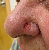
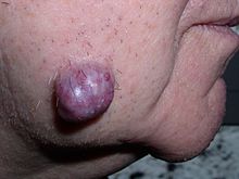
- سرطانة الخلايا القاعدية
- Acanthoma fissuratum (granuloma fissuratum، spectacle frame acanthoma)
- Acrospiroma (clear cell hidradenoma، dermal duct tumor، hidroacanthoma simplex، nodular hidradenoma، poroma)
- تقران سفعي (senile keratosis، solar keratosis)
- سرطانة حرشفية الخلايا (pseudoglandular squamous cell carcinoma)
- Aggressive digital papillary adenocarcinoma (digital papillary adenocarcinoma، papillary adenoma)
سرطانة الخلايا القاعدية - سرطانة الغدة المفترزة
- وحمة مفترزة
- Arsenical keratosis
- تقران سفعي
- التهاب الحشفة البلازماوي (balanoposthitis chronica circumscripta plasmacellularis، balanitis circumscripta plasmacellularis، plasma cell balanitis، plasma cell vulvitis، vulvitis circumscripta plasmacellularis، Zoon's balanitis، Zoon's erythroplasia، Zoon's vulvitis)
- سرطانة الخلايا القاعدية
- Basaloid follicular hamartoma
- سرطانة حرشفية الخلايا
- متلازمة بيرت-هوغ-دوبي
- سرطان الجلد حرشفي الخلايا (squamous cell carcinoma in situ)
- Brooke–Fordyce syndrome
- Ceruminoma
- سرطانة الخلايا القاعدية (morpheaform basal cell carcinoma، morphoeic basal cell carcinoma)
- Ciliated cyst of the vulva (cutaneous Müllerian cyst، paramesonephric mucinous cyst of the vulva)
- Clear cell acanthoma (acanthome cellules claires of Degos and Civatte، Degos acanthoma، pale cell acanthoma)
- سرطانة حرشفية الخلايا (clear cell carcinoma of the skin)
- Chronic scar keratosis (chronic cicatrix keratosis)
- تقران مثي
- تقران مثي (basal cell papilloma، solid seborrheic keratosis)
- متلازمة كاودن (Cowden's disease، multiple hamartoma syndrome)
- Cutaneous ciliated cyst
- Cutaneous columnar cyst
- قرن جلدي (Cornu cutaneum)
- سرطانة الخلايا القاعدية
- ورم جلدي أسطواني الخلايا (cylindroma)
- Dermatosis papulosa nigra
- ورم ظهاري شعري ليفي
- Dilated pore (dilated pore of Winer)

سرطان الجلد حرشفي الخلايا - Eccrine carcinoma (syringoid carcinoma)
- Eccrine nevus
- كيسة بشرانية (epidermal inclusion cyst، epidermoid cyst، infundibular cyst، keratin cyst)
- متلازمة الوحمة البشروية (Feuerstein and Mims syndrome، Solomon's syndrome)
- Epidermolytic acanthoma
- سرطانة ثؤلولية (Ackerman tumor، carcinoma cuniculatum)
- Eruptive vellus hair cyst
- سرطان الجلد حرشفي الخلايا
- داء باجيت خارج الثدي
- سرطانة الخلايا القاعدية
- سرطانة الخلايا القاعدية
- ورم جريبي ليفي
- Follicular hybrid cyst (Hybrid cyst)
- Folliculosebaceous-apocrine hamartoma (follicular-apocrine hamartoma)
- Folliculosebaceous cystic hamartoma
- الشوكوم القرني (generalized eruptive keratoacanthoma of Grzybowski)
- Giant solitary trichoepithelioma
- Hidradenoma
Hidradenoma - Hidradenocarcinoma
- Hidrocystoma (cystadenoma، Moll's gland cyst، sudoriferous cyst)
- تقران هيدروكربوني (pitch keratosis، tar keratosis، tar wart)
- فرط التقرن (Flegel's disease)
- فرط التقرن
- تقران سفعي
- داء السمكية القنفذي (ichthyosis hystrix gravior type Lambert، porcupine man، systematized verrucous nevus)
- داء السمكية القنفذي
- سرطانة الخلايا القاعدية
- Inflammatory linear verrucous epidermal nevus
- تقران مثي
- تقران مثي (basosquamous cell acanthoma، inflamed seborrheic keratosis)
- Isthmicoma (infundibuloma، tumor of the follicular infundibulum)
- ابيضاض وحيدي نقوي طفولي
- Keratin implantation cyst
- الشوكوم القرني
- الشوكوم القرني
- Large cell acanthoma
- تقران سفعي
- حزاز مسطح (benign lichenoid keratosis، lichen planus-like keratosis، solitary lichen planus، solitary lichenoid keratosis)
- Linear verrucous epidermal nevus (linear epidermal nevus، verrucous epidermal nevus)
- ورم نهايات الغدة العرقية الخبيث (malignant poroma، porocarcinoma، spiradenocarcinoma)
- ورم مختلط خبيث (malignant chondroid syringoma)
- Malignant trichilemmal cyst
- Mantleoma
- قرحة مارجولين
- تقران مثي (pigmented seborrheic keratosis)
- سرطانة خلية مركل (cutaneous apudoma، primary neuroendocrine carcinoma of the skin، primary small cell carcinoma of the skin، trabecular carcinoma of the skin)
- Microcystic adnexal carcinoma (sclerosing sweat duct carcinoma)
- سرطانة الخلايا القاعدية
- Milia en plaque
- دخينة

دخينة - Mixed tumor (chondroid syringoma)
- سرطانة مخاطية
- وحمة مخاطية (nevus mucinosus)
- متلازمة موير توري
- Multiple familial trichoepithelioma (Brooke–Spiegler syndrome، epithelioma adenoides cysticum)
- الشوكوم القرني (Ferguson–Smith syndrome، Ferguson-Smith type of multiple self-healing keratoacanthomas، multiple keratoacanthomas of the Ferguson–Smith type)
- Multiple minute digitate hyperkeratosis (digitate keratoses، disseminated spiked hyperkeratosis، familial disseminated piliform hyperkeratosis، minute aggregate keratosis)
- متلازمة سرطانة الخلية القاعدية الوحمانية (basal cell nevus syndrome، Gorlin syndrome، Gorlin–Goltz syndrome)
- Nevus comedonicus (comedo nevus)
- Nevus comedonicus syndrome
- Nevus sebaceous (nevus sebaceous of Jadassohn، organoid nevus)
- Nevus unius lateris
- سرطانة الخلايا القاعدية (classic basal cell carcinoma)
- داء باجيت في الثدي
- Papillary eccrine adenoma (tubular apocrine adenoma)
- ورم غدي عرقي حليمي (hidradenoma papilliferum)
- Papillomatosis cutis carcinoides (Gottron's carcinoid papillomatosis، papillomatosis cutis carcinoides of Gottron–Eisenlohr)
- Patch blue nevus (acquired dermal melanocytosis، dermal melanocyte hamartoma)
- ورم ليفي محيط بالجريب
- Phakomatosis pigmentokeratotica
- تقران سفعي
- سرطانة الخلايا القاعدية
- Pigmented hairy epidermal nevus syndrome
- Pilar sheath acanthoma
- ناسور شعري (Barber's interdigital pilonidal sinus، pilonidal cyst، pilonidal disease)
- سرطانة الخلايا القاعدية
- سرطانة الخلايا القاعدية
- Primary cutaneous adenoid cystic carcinoma
- Proliferating epidermoid cyst (proliferating epithelial cyst)
- Proliferating trichilemmal cyst (pilar tumor، proliferating follicular cystic neoplasm، proliferating pilar tumor، proliferating trichilemmal tumor)
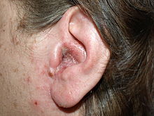
- Pseudocyst of the auricle (auricular endochondrial pseudocyst، cystic chondromalacia، endochondral pseudocyst، intracartilaginous cyst)
- التهاب الحشفة التقرني الظهاري الكاذب والصخري الشكل
- PUVA keratosis
- متلازمة راسموسن
- Reactional keratosis
- تقران مثي (adenoid seborrheic keratosis)
- سرطانة الخلايا القاعدية (Jacobi ulcer)
- متلازمة شيميلبنينغ (Schimmelpenning–Feuerstein–Mims syndrome)
- ورم ظهاري زهمي (sebaceous epithelioma)
- ورم غدي دهني
- سرطانة زهمية
- فرط تنسج دهني
- Sebaceous nevus syndrome
- Seboacanthoma
- تقران مثي (seborrheic verruca، senile wart)
- تقران مثي
- سرطانة حرشفية الخلايا
- الشوكوم القرني (subungual keratoacanthoma)
- ورم ظهاري شعري منفرد
- سرطانة مغزلية الخلايا (spindle cell carcinoma)
- ورم الغدة العرقية
- سرطانة حرشفية الخلايا

سرطانة حرشفية الخلايا - ورم كيسي دهني متعدد (epidermal polycystic disease، sebocystomatosis)
- ورم كيسي دهني بسيط (simple sebaceous duct cyst، solitary steatocystoma)
- تقران مثي (digitate seborrheic keratosis، hyperkeratotic seborrheic keratosis، keratosis alba، serrated seborrheic keratosis، verrucous seborrheic keratosis)
- سرطانة الخلايا القاعدية (superficial multicentric basal cell carcinoma)
- Syringadenoma papilliferum (syringocystadenoma papilliferum)
- Syringofibroadenoma (acrosyringeal nevus of Weedon and Lewis)
- غدوم عرقي (ورم غدي عرقي)
- Systematized epidermal nevus
- تقران حراري
- Trichilemmal carcinoma
- كيسة شعرية (isthmus-catagen cyst، pilar cyst)
- Trichilemmoma
- Trichoadenoma (trichoadenoma of Nikolowski)
- ورم قرص الشعرة
- ورم قرص الشعرة
- Trichodiscoma
- Trichofolliculoma
- Unilateral palmoplantar verrucous nevus
- ورم الإحليل
- سرطان ثؤلولي
- Verrucous cyst (cystic papilloma)
- ثؤلول
- Warty dyskeratoma (isolated dyskeratosis follicularis)
- Waxy keratosis of childhood (kerinokeratosis papulosa)
- التهاب الحشفة البلازماوي
- Zosteriform speckled lentiginous nevus


أمراض حمامية[عدل]

حمامىs are reactive skin conditions in which there is ابيضاض (طب) redness.[1][9]
- Erythema annulare centrifugum (deep gyrate erythema، erythema perstans، palpable migrating erythema، superficial gyrate erythema)
- Erythema gyratum repens (Gammel's disease)
- Erythema migrans (erythema chronicum migrans)
- حمامى متعددة الأشكال
- Erythema multiforme minor (herpes simplex-associated erythema multiforme)
- حمامى راحية
- Generalized erythema
- Necrolytic acral erythema
- Necrolytic migratory erythema (glucagonoma syndrome)
الجلادات الوراثية[عدل]
جلاد وراثي are توريث (أحياء) genetic skin conditions often grouped into three categories: كروموسوم، single جين، and polygenetic.[64][65]
- 18q deletion syndrome
- التهاب الجلد النهائي المعوي
- Acrogeria (Gottron syndrome)
- Acrokeratosis verruciformis (acrokeratosis verruciformis of Hopf)
- متلازمة آدامز أوليفر
- متلازمة الإبهام المقرب
- Albright's hereditary osteodystrophy
- متلازمة أنجلمان
- متلازمة أبير (acrocephalosyndactyly)
- Arthrogryposis–renal dysfunction–cholestasis syndrome
- رنح توسع الشعيرات (Louis–Bar syndrome)
- Atrichia with papular lesions (papular atrichia)
- ضمور الجلد الدودي الشكل (acne vermoulante، acne vermoulanti، atrophoderma reticulata symmetrica faciei، atrophoderma reticulatum، atrophoderma vermiculata، atrophoderma vermiculatum، atrophodermia reticulata symmetrica faciei، atrophodermia ulerythematosa، atrophodermie vermiculée des joues avec kératoses folliculaires، folliculitis ulerythema reticulata، folliculitis ulerythematous reticulata، folliculitis ulerythemosa، honeycomb atrophy، ulerythema acneforme، ulerythema acneiforme)
- متلازمة متعدد الغدد الصم بالمناعة الذاتية النوع الأول
- Bart syndrome
- Bazex–Dupré–Christol syndrome (Bazex syndrome، follicular atrophoderma and basal cell cacinomas)
- Beare–Stevenson cutis gyrata syndrome
- متلازمة بلوم (Bloom–Torre–Machacek syndrome)
- متلازمة الوحمة الزرقاء المطاطية الفقاعية
- حثل كبريتي شعري
- متلازمة كانتو
- Cardio-facio-cutaneous syndrome (cardiofaciocutaneous syndrome)
- نقص تنسج الغضروف والشعر (McKusick type metaphyseal chondrodysplasia)
- Cerebral dysgenesis–neuropathy–ichthyosis–keratoderma syndrome
- Childhood tumor syndrome
- خلل التنسج الغضروفي المنقط
- انحلال البشرة الفقاعي الموصلي
- Craniosynostosis–anal anomalies–porokeratosis syndrome
- متلازمة كوكاين
- Colobomas of the eye–heart defects–ichthyosiform dermatosis–mental retardation–ear defects syndrome (CHIME syndrome، Zunich neuroectodermal syndrome، Zunich–Kaye syndrome)
- متلازمة تشايلد (CHILD syndrome)
- متلازمة كونرادي هيونرمان (Conradi–Hünermann–Happle syndrome، Happle syndrome، X-linked dominant chondrodysplasia punctata)
- متلازمة كوستيلو
- متلازمة كرونخيت-كندا
- متلازمة كروزن
- Cutis verticis gyrata
- داء دارييه (Darier–White disease، dyskeratosis follicularis، keratosis follicularis)

Linear Darier disease - DeSanctis–Cacchione syndrome
- Disseminated superficial actinic porokeratosis
- تقران منخرب
- عوز كيناز الدوليكول
- انحلال البشرة الفقاعي الحثلي
- خلل التقرن الخلقي (Zinsser–Cole–Engman syndrome)
- انحلال البشرة الفقاعي الحثلي
- خلل التنسج الأديمي الظاهر
- Ectodermal dysplasia with corkscrew hairs
- متلازمة انعدام الأصابع وخلل التنسج الأديمي (EEC syndrome، split hand–split foot–ectodermal dysplasia–cleft syndrome)
- Epidermolysis bullosa herpetiformis (Dowling–Meara epidermolysis bullosa simplex)
- Epidermolysis bullosa simplex
- Epidermolysis bullosa simplex of Ogna
- Epidermolysis bullosa simplex with mottled pigmentation
- Epidermolysis bullosa simplex with muscular dystrophy
- Epidermolytic hyperkeratosis (bullous congenital ichthyosiform erythroderma، bullous ichthyosiform erythroderma)
- تقرن الجلد الاحمراري مع رنح (Giroux–Barbeau syndrome)
- مرض هيلي- هيلي (familial benign pemphigus، Hailey–Hailey disease)
- متلازمة فانكوني (familial pancytopenia، familial panmyelophthisis)
- خلل التنسج الليفي المعظم المترقي
- نقص تنسج الجلد البؤري (Goltz syndrome)
- Follicular atrophoderma
- Franceschetti–Klein syndrome (mandibulofacial dysostosis)
- متلازمة غاردنر (familial colorectal polyposis)
- Gastrocutaneous syndrome
- انحلال البشرة الفقاعي الموصلي
- Generalized epidermolysis bullosa simplex (Koebner variant of generalized epidermolysis bullosa simplex)
- Generalized trichoepithelioma
- اعتلال عصبي عملاق المحوار مع شعر مجعد
- Gingival fibromatosis with hypertrichosis
- Haber syndrome
- متلازمة هاليرمان-سترايف
- الوليد المهرج (harlequin baby، harlequin fetus، harlequin ichthyosis، ichthyosis congenita، ichthyosis congenita gravior)
- Hay–Wells syndrome (AEC syndrome، ankyloblepharon filiforme adnatum–ectodermal dysplasia–cleft palate syndrome، ankyloblepharon–ectodermal defects–cleft lip and palate syndrome، ankyloblepharon–ectodermal dysplasia–clefting syndrome)
- Hereditary sclerosing poikiloderma

تقرن الجريبات الشعرية - التباين في لون العين
- Holocarboxylase synthetase deficiency
- خلل التنسج الأديمي الظاهر ناقص التعرق (anhidrotic ectodermal dysplasia، Christ–Siemens–Touraine syndrome)
- Hypotrichosis–acro-osteolysis–onychogryphosis–palmoplantar keratoderma–periodontitis syndrome
- Hypotrichosis–lymphedema–telangiectasia syndrome
- حثل كبريتي شعري (IBIDS syndrome، sulfur-deficient brittle hair syndrome، Tay's syndrome، trichothiodystrophy، trichothiodystrophy with ichthyosis)
- سماك سيمنز الفقاعي (ichthyosis exfoliativa)
- Ichthyosis follicularis (ichthyosis follicularis with alopecia and photophobia syndrome)
- Ichthyosis linearis circumflexa
- متلازمة سماك الولادة المبكرة
- سماك شائع (autosomal dominant ichthyosis، ichthyosis simplex)
- Ichthyosis with confetti
- Neonatal ichthyosis–sclerosing cholangitis syndrome (ichthyosis–sclerosing cholangitis syndrome، NISCH syndrome)
- سلس الصباغ المنقص التلون (hypomelanosis of Ito)
- متلازمة إبكس
- Jaffe–Campanacci syndrome
- متلازمة جوهانسون-بليزارد
- Johnson–McMillin syndrome
- متلازمة جيوبرت
- انحلال البشرة الفقاعي الموصلي
- Junctional epidermolysis bullosa gravis (epidermolysis bullosa letalis، Herlitz disease، Herlitz epidermolysis bullosa، Herlitz syndrome، lethal junctional epidermolysis bullosa)
- Junctional epidermolysis bullosa with pyloric atresia
- متلازمة كابوكي (Kabuki makeup syndrome، Niikawa–Kuroki syndrome)
- حمامى الشتاء الحالة للطبقة القرنية (erythrokeratolysis hiemalis، Oudtshoorn disease، Oudtshoorn skin)
- تقران جريبي شوكي صالع (Siemens-1 syndrome)
- Keratosis linearis with ichthyosis congenital and sclerosing keratoderma syndrome
- تقران شعري أحمر ضموري وجهي (folliculitis rubra، keratosis pilaris rubra atrophicans faciei، lichen pilare، lichen pilaire ou xerodermie pilaire symmetrique de la face'، ulerythema ophryogenes، xerodermi pilaire symmetrique de la face)
- تقرن الجريبات الشعرية
- Kindler syndrome (acrokeratotic poikiloderma، bullous acrokeratotic poikiloderma of Kindler and Weary، congenital poikiloderma with blisters and keratoses، congenital poikiloderma with bullae and progressive cutaneous atrophy، hereditary acrokeratotic poikiloderma، hyperkeratosis–hyperpigmentation syndrome، Weary–Kindler syndrome)
- متلازمة كلاينفيلتر
- متلازمة كليبل فيل
- سماك صفاحي (collodion baby)
- متلازمة ليجيوس (neurofibromatosis type 1-like syndrome)
- Lelis syndrome
- Lenz–Majewski syndrome
- Leschke syndrome
- Lethal acantholytic epidermolysis bullosa
- Lhermitte–Duclos disease
- Linear and whorled nevoid hypermelanosis (linear nevoid hyperpigmentation، progressive cribriform and zosteriform hyperpigmentation، reticulate and zosteriform hyperpigmentation، reticulate hyperpigmentation of Iijima and Naito and Uyeno، zebra-like hyperpigmentation in whorls and streaks، zebra-line hyperpigmentation)
- Linear Darier disease (acantholytic dyskeratotic epidermal nevus)
- تقران منخرب
- Localized epidermolysis bullosa simplex (Weber–Cockayne syndrome، Weber–Cockayne variant of generalized epidermolysis bullosa simplex)
- Mandibuloacral dysplasia
- متلازمة مارينسكو شوغرن
- متلازمة ماكيون أولبرايت
- McCusick syndrome
- شياخ
- Microphthalmia–dermal aplasia–sclerocornea syndrome
- انحلال البشرة الفقاعي الموصلي (nonlethal junctional epidermolysis bullosa)
- متلازمة ميلاس
- متلازمة نونان ذات التصبغات المتعددة (cardiocutaneous syndrome، Gorlin syndrome II، lentiginosis profusa syndrome، LEOPARD syndrome، progressive cardiomyopathic lentiginosis)
- Multiple pterygium syndrome
- عوز السلفاتاز المتعدد (Austin disease، mucosulfatidosis)
- Naegeli–Franceschetti–Jadassohn syndrome (chromatophore nevus of Naegeli)
- Netherton syndrome
- ورم عصبي ليفي من النوع الأول (von Recklinghausen's disease)

ورم عصبي ليفي من النوع الأول - Neurofibromatosis type 3 (neurofibromatosis mixed type)
- Neurofibromatosis type 4 (neurofibromatosis variant type)
- مرض تخزين الدهون المحايد (Dorfman–Chanarin syndrome)
- Nonbullous congenital ichthyosiform erythroderma (congenital ichthyosiform erythroderma)
- متلازمة نوونان
- Oculocerebrocutaneous syndrome (Delleman–Oorthuys syndrome)
- Oculodentodigital dysplasia
- Odonto–Tricho–Ungual–Digital–Palmar syndrome
- Oliver–McFarlane syndrome
- متلازمة فموية إصبعية وجهية نمط 1
- ثخن الجلد وتعظم السمحاق (idiopathic hypertrophic osteoathorpathy، Touraine–Solente–Gole syndrome)
- متلازمة تقشير الجلد (acral peeling skin syndrome، continual peeling skin syndrome، familial continual skin peeling، idiopathic deciduous skin، keratolysis exfoliativa congenita)
- متلازمة فايفر
- حثل كبريتي شعري
- Pityriasis rotunda (pityriasis circinata، tinea circinata)
- Plate-like osteoma cutis
- تقران منخرب (classic porokeratosis، porokeratosis of Mibelli)
- متلازمة POEMS (Crow–Fukase syndrome)
- خلل التنسج الليفي متعدد العظام (Albright's disease)
- متلازمة الظفرة المابضية
- تقران منخرب
- تقران منخرب
- متلازمة برادر- فيلي
- شياخ (Hutchinson–Gilford progeria syndrome، Hutchinson–Gilford syndrome، progeria syndrome)
- Progressive osseous heteroplasia
- تقرن الجلد الاحمراري المتناظر المترق (erythrokeratodermia progressiva symmetrica)
- متلازمة بروتيوس
- متلازمة بروتيوس الشبيهة
- تقران منخرب
- Rapp–Hodgkin syndrome (Rapp–Hodgkin ectodermal dysplasia syndrome)
- انحلال البشرة الفقاعي الحثلي (Hallopeau–Siemens variant of epidermolysis bullosa، Hallopeau–Siemens disease)
- داء ريفسام (heredopathia atactica polyneuritiformis، phytanic acid storage disease)
- جلاد انحلال الأشواك الخطي الناكس
- Restrictive dermopathy

سماك مرتبط بX - Rhizomelic chondrodysplasia punctata (autosomal recessive chondrodysplasia punctata type 1، chondrodystrophia calcificans punctata، peroxisomal biogenesis disorder complementation group 11)
- Rombo syndrome
- متلازمة روثموند تومسون (poikiloderma congenitale)
- Rud syndrome
- Say syndrome
- Scalp–ear–nipple syndrome (Finlay–Marks syndrome)
- Schindler disease (Kanzaki disease، alpha-N-acetylgalactosaminidase deficiency)
- Schinzel–Giedion syndrome
- تقرن الجلد الراحي الأخمصي (Huriez syndrome، palmoplantar keratoderma with scleroatrophy، palmoplantar keratoderma with sclerodactyly، scleroatrophic and keratotic dermatosis of the limbs، sclerotylosis)
- ورم عصبي ليفي من النوع الأول
- Senter syndrome (Desmons' syndrome)
- Shabbir syndrome (laryngo–onycho–cutaneous syndrome)
- متلازمة سيلفر-روسال
- متلازمة شوغرن لارسون
- Skin fragility syndrome (plakophilin 1 deficiency)
- متلازمة سميث-ليملي-أوبيتز
- متلازمة ستيرج ويبر
- Supernumerary nipples–uropathies–Becker's nevus syndrome
- Terminal osseous dysplasia with pigmentary defects
- Tooth and nail syndrome (hypodontia with nail dysgenesis، Witkop syndrome)
- Townes–Brocks syndrome
- Transient bullous dermolysis of the newborn

جفاف الجلد المصطبغ - متلازمة تريتشر كولينز (Treacher Collins–Franceschetti syndrome)
- Tricho–dento–osseous syndrome
- Tricho–rhino–phalangeal syndrome
- تصلب حدبي (Bourneville disease، epiloia)
- متلازمة تيرنر
- Ulnar–mammary syndrome
- Van Der Woude syndrome
- داء فون هيبل - لينداو
- متلازمة واطسون
- متلازمة فيرنر (adult progeria)
- Westerhof syndrome
- متلازمة فريمان-شيلدون (craniocarpotarsal syndrome، distal arthrogryposis type 2، Freeman–Sheldon syndrome، Windmill–Vane–Hand syndrome)
- Wilson–Turner syndrome
- متلازمة وولف - هيرشيرون (4p- syndrome)
- سماك مرتبط بX (steroid sulfatase deficiency، X-linked recessive ichthyosis)
- X-linked recessive chondrodysplasia punctata
- جفاف الجلد المصطبغ (Cockayne syndrome complex)
- متلازمة XXYY
- متلازمة زيمرمان–لاباند


أمراض مرتبطة بالعدوى[عدل]
Infection-related cutaneous conditions may be caused by بكتيريا، فطر، خميرة، فيروسes، and/or تطفل.[26][66]
أمراض مرتبطة بعدوى بكتيرية[عدل]
Bacterium-related cutaneous conditions often have distinct علم التشكل (أحياء) characteristics that may be an indication of a generalized systemic process or simply an isolated superficial infection.[66][67]
- عدوى الآيروموناس
- حمى لدغة القرادة الإفريقية
- American tick bite fever (Rickettsia parkeri infection)
- Arcanobacterium haemolyticum infection
- ورام وعائي عصوي
- بجل (endemic syphilis)
- Blastomycosis-like pyoderma (pyoderma vegetans)
- تبثر التهاب الإصبع القاصي
- Botryomycosis
- داء بريل زينسر
- داء البروسيلات (Bang's disease، Malta fever، undulant fever)
- Bullous impetigo
- مرض خدش القطة (cat scratch fever، English–Wear infection، inoculation lymphoreticulosis، subacute regional lymphadenitis)
- التهاب الهلل

التهاب الهلل - قرحة صلبة
- قريح (soft chancre، ulcus molle)
- داء المتدثرات
- التهاب الأوعية اللمفاوية المزمن
- الحمرة
- Chronic undermining burrowing ulcers (Meleney gangrene)
- Chromobacteriosis infection
- ورم لقمي مسطح
- Cutaneous actinomycosis
- جمرة خبيثة
- Cutaneous C. diphtheriae infection (Barcoo rot، diphtheric desert sore، septic sore، Veldt sore)
- Cutaneous group B streptococcal infection
- Cutaneous Pasteurella hemolytica infection
- Cutaneous Streptococcus iniae infection
- Dermatitis gangrenosa (gangrene of the skin)
- إكثيمة
- Ecthyma gangrenosum
- Ehrlichiosis ewingii infection
- داء الفيل المتوطن
- Endemic typhus (murine typhus)
- تيفوس وبائي (epidemic louse-borne typhus)
- الحمرة (ignis sacer، Saint Anthony's fire)
- Erysipeloid of Rosenbach
- حمامى هامشية
- وذح
- التهاب الأذن الخارجية (otitis externa، swimmer's ear)
التهاب الأذن الخارجية - داحس
- Flea-borne spotted fever
- Flinders Island spotted fever
- Flying squirrel typhus
- التهاب جريبي
- غنغرينة فورنير (Fournier gangrene of the penis or scrotum)
- الدمل (boil)
- غنغرينا غازية (clostridial myonecrosis، myonecrosis)
- رعام (equinia، farcy، malleus)
- Gonococcemia (arthritis–dermatosis syndrome، disseminated gonococcal infection)
- سيلان (مرض) (clap)
- Gram-negative folliculitis
- Gram-negative toe web infection
- داء الدونوفانيات (Donovanosis، granuloma genitoinguinale، granuloma inguinale tropicum، granuloma venereum، granuloma venereum genitoinguinale، lupoid form of groin ulceration، serpiginous ulceration of the groin، ulcerating granuloma of the pudendum، ulcerating sclerosing granuloma)
- Green nail syndrome
- Group JK Corynebacterium sepsis
- Haemophilus influenzae cellulitis
- Helicobacter cellulitis
- Hospital furunculosis
- طفح الحمام الحار (Pseudomonas aeruginosa folliculitis)
- عدوى المحببات البشرية بالإيرليخية
- المرض الايرليخي البشري ذات التوجه الاحادي للخلايا
- قوباء
- Japanese spotted fever
- داء البريميات (Fort Bragg fever، pretibial fever، Weil's disease)
- داء الليستريات
- ذباح لودفيغ
- تينة ذأبانية
- داء لايم (Afzelius' disease، Lyme borreliosis)
- دبل مناخي (climatic bubo، Durand–Nicolas–Favre disease، lymphogranuloma inguinale، poradenitis inguinale، strumous bubo)
- تلين لويحي (malacoplakia)
- حمى برعمية (Boutonneuse fever)
- راعوم (Whitmore's disease)
- مرض المكورات السحائية
- القرادة الجنوبية-مرتبط بمرض الطفح
- مفطورة
- مرض أكل اللحم (flesh-eating bacteria syndrome)
- Neonatal toxic shock-like exanthematous disease
- داء النوكارديات
- Noma neonatorum
- North Asian tick typhus
- رمد وليدي
الحمرة - داء كاريون (Carrion's disease)
- داء الباستوريلات
- Perianal cellulitis (perineal dermatitis، streptococcal perianal disease)
- خراج سني
- زهري مستوطن
- Pitted keratolysis (keratolysis plantare sulcatum، keratoma plantare sulcatum، ringed keratolysis)
- طاعون
- التهاب الجلد الأولي بالمكورات البنية
- Pseudomonal pyoderma
- Pseudomonas hot-foot syndrome
- تقرح حول ظفري
- التهاب عضلات صديدي
- حمى كيو
- Queensland tick typhus
- حمى عضة الجرذ
- Recurrent toxin-mediated perineal erythema
- ورم صلب أنفي
- Rickettsia aeschlimannii infection
- وقس ريكتسي
- حمى الجبال الصخرية المبقعة
- قصبة الساق السيفية (anterior tibial bowing)
- أنف سرجي
- داء السلمونيلات
- حمى قرمزية
- تيفوس أكالي (Tsutsugamushi fever)
- زحار
- متلازمة الجلد المسموط العنقودية (pemphigus neonatorum، Ritter's disease)
- Streptococcal intertrigo
- Superficial pustular folliculitis (impetigo of Bockhart، superficial folliculitis)
- سيكوز (barber's itch، sycosis barbae)
- Syphilid
- زهري (lues)
- Tick-borne lymphadenopathy
- متلازمة الصدمة التسممية (streptococcal toxic shock syndrome، streptococcal toxic shock-like syndrome، toxic streptococcal syndrome)
- حمى الخنادق (five-day fever، quintan fever، urban trench fever)
- قرحة مدارية (Aden ulcer، jungle rot، Malabar ulcer، tropical phagedena)
- داء توليري (deer fly fever، Ohara's disease، Pahvant Valley plague، rabbit fever)
- داء كاريون
- Vibrio vulnificus infection
- الداء العليقي (bouba، frambösie، parangi، pian)


أمراض مرتبطة بعدوى المتفطرة[عدل]
Mycobacterium-related cutaneous conditions are caused by متفطرة infections.[66][68]
- Aquarium granuloma (fish-tank granuloma، swimming-pool granuloma)
- جذام ورمي حدي
- جذام حدي
- جذام شبه درني حدي
- قرحة بورولي (Bairnsdale ulcer، Searl ulcer، Searle's ulcer)

قرحة بورولي - حمامى جاسية (Bazin disease)
- جذام شبه نسيجي
- جذام ورمي
- جذام (Hansen's disease)
- حزاز خنازيري (tuberculosis cutis lichenoides)
- ذئبة شائعة (tuberculosis luposa)
- سل دخني (disseminated tuberculosis، tuberculosis cutis acuta generalisata، tuberculosis cutis disseminata)
- عدوى المتفطرة الطيرية داخل الخلوية
- Mycobacterium haemophilum infection
- متفطرة كنساس
- طفحة سلية حطاطية نخرية
- Primary inoculation tuberculosis (cutaneous primary complex، primary tuberculous complex، tuberculous chancre)
- Rapid-growing Mycobacterium infection
- Scrofuloderma (tuberculosis cutis colliquativa)
- سل الفوهات (acute tuberculous ulcer، orificial tuberculosis)
- سل ثؤلولي (lupus verrucosus، prosector's wart، warty tuberculosis)
- Tuberculous cellulitis
- Tuberculous gumma (metastatic tuberculous abscess، metastatic tuberculous ulcer)
- جذام شبه درني

أمراض فطرية[عدل]
Mycosis-related cutaneous conditions are caused by فطر or خميرةs، and may present as either a superficial or deep عدوى of the skin، hair، and/or nails.[66]
- داء النوسجات الأفريقي
- Alternariosis
- داء المبيضات (iatrogenic candidiasis)
- بيصرة سوداء
- Candidal intertrigo
- Candidal onychomycosis
- تقرح حول ظفري
- عدوى مهبلية بالخميرة
- طفحة المبيضات
- فطار اصطباغي (chromomycosis، cladosporiosis، Fonseca's disease، Pedroso's disease، phaeosporotrichosis، verrucous dermatitis)
- Chronic mucocutaneous candidiasis
- حمى الصحراء (California disease، desert rheumatism، San Joaquin Valley fever، valley fever)
- Congenital cutaneous candidiasis
- داء المستخفيات
- Dermatophytid
- داء المبيضات
- Disseminated coccidioidomycosis (coccidioidal granuloma)
- Distal subungual onychomycosis
- Entomophthoromycosis
- Erosio interdigitalis blastomycetica

سعفة قرعية - سعفة قرعية
- التهاب الأجربة الفطري (majocchi granuloma)
- Fusariosis
- داء التيربيات
- Granuloma gluteale infantum
- داء النوسجات (cave disease، Darling's disease، Ohio Valley disease، reticuloendotheliosis)
- Hyalohyphomycosis
- شهدة
- Lobomycosis (keloidal blastomycosis، lacaziosis، Lobo's disease)
- فطر عفني
- Mycetoma (Madura foot، maduromycosis)
- فطار برعمي (blastomycetic dermatitis، blastomycosis، Gilchrist's disease)
- التهاب الظفر الفطري (dermatophytic onychomycosis، ringworm of the nail، tinea unguium)
- داء المبيضات الفموي (thrush)
- فطار أذني
- داء المبيضات
- التهاب الشفة الزاوي (angular cheilitis)
- داء الفطار العبسي
- بيصرة (trichosporosis)
- Pityrosporum folliculitis
- Primary cutaneous aspergillosis
- Primary cutaneous coccidioidomycosis
- داء النوسجات الجلدي الأولي
- Primary pulmonary coccidioidomycosis
- داء النوسجات الرئوي الأولي
- داء النوسجات المنتثر المترق
- Proximal subungual onychomycosis
- Rhinosporidiosis
- الفطار نظير الكرواني (Brazilian blastomycosis، paracoccidioidal granuloma، paracoccidioidomycosis)
- داء الشعريات المبوغة (rose-gardener's disease)
- Systemic candidiasis
- سعفة الرأس (barber's itch، ringworm of the beard، tinea sycosis)

سعفة الرأس - سعفة الرأس (herpes tonsurans، ringworm of the hair، ringworm of the scalp، scalp ringworm، tinea tonsurans)
- سعفة الجسم (ringworm، tinea circinata، tinea glabrosa)
- سعفة الجسم
- سعفة ساقية (crotch itch، eczema marginatum، gym itch، jock itch، ringworm of the groin)
- Tinea faciei
- Tinea imbricata (tokelau)
- Tinea incognito
- سعفة اليد
- Tinea nigra (superficial phaeohyphomycosis، tinea nigra palmaris et plantaris)
- سعفة القدم (athlete's foot، ringworm of the foot)
- نخالية مبرقشة (dermatomycosis furfuracea، pityriasis versicolor، tinea flava)
- بيصرة بيضاء
- White superficial onychomycosis
- فطار عفني (phycomycosis)


الغزو الطفيلي واللسعات واللدغات[عدل]
Parasitic infestations، stings، and bites in humans are caused by several groups of organisms belonging to the following شعبة (تصنيف): حلقيات، مفصليات الأرجل، مرجانيات، حبليات، لاسعات، زراقم، شوكيات الجلد، ديدان أسطوانية، ديدان مسطحة، and أولي (كائن).[66][69]
- عدوى الشوكميبة
- داء الأميبات الجلدي
- Ant sting
- لدغات العنكبوت
- حكة الخبازين
- Balamuthia infection
- عدوى بطفيليات بق الفراش (bedbug bite، cimicosis)
- لسعة النحل
- Blister beetle dermatitis
- خنفساء بومباردييه
- كثيرات الأشعار
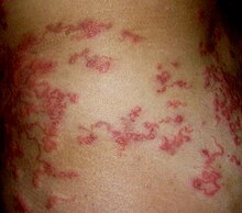
داء هجرة اليرقات الجلدي - Centipede bite
- Cheyletiella dermatitis
- داء الخطماوات
- Coolie itch
- حكة تراب جوز الهند
- التهاب جلدي مرجاني
- داء هجرة اليرقات الجلدي (cutaneous larva migrans)
- داء الليشمانيات الجلدي (Aleppo boil، Baghdad boil، bay sore، Biskra button، Chiclero ulcer، Delhi boil، Kandahar sore، Lahore sore، leishmaniasis tropica، oriental sore، pian bois، uta)
- Cysticercosis cutis
- Demodex mite bite
- Dogger Bank itch
- داء التنينات (dracontiasis، guinea worm disease، Medina worm)
- داء المشوكات (hydatid disease)
- داء الفيل (elephantiasis arabum)
- داء كلابية الذنب
- داء السرميات (oxyuriasis، pinworm infection، seatworm infection)
- Erisipela de la costa
- التهاب الجلد الوسادي الريشي
- لدغات العنكبوت
- داء القمقاميات
- داء الفغميات (larva migrans profundus)
- العث السمين (barley itch، mattress itch، prairie itch، straw itch)
- Grocer's itch
- عدوى قمل الرأس (cooties، pediculosis capitis)
- دودة شصية (دودة الانسيلوستوما) (ancylostomiasis، ground itch، necatoriasis، uncinariasis)
- Human trypanosomiasis
- Hydroid dermatitis
- متلازمة الإروكانجي
- Jellyfish dermatitis
- حكة الطبوع
- Larva currens
- تسمم بزعاف الزوع (widow spider bite)
- علقيات
- داء كلابية الذنب
- Lepidopterism (Caripito itch، caterpillar dermatitis، moth dermatitis)
- Lizard bite
- داء كلابية الذنب
- داء اللوائيات (Calabar swelling، fugitive swelling، loa loa، tropical swelling)
- Loxoscelism (brown recluse spider bite، necrotic cutaneous loxoscelism)
- Mal morando
- Millipede burn
- بعوضيات
- داء الليشمانيات (espundia، leishmaniasis americana)
- نغف
- التهاب الجلد بحشرة البيدروس (Kenya fly dermatitis، Nairobi eye)
- التهاب الجلد النيماتودي
- جرب (crusted scabies)
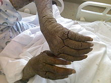
جرب - داء كلابية الذنب
- Ophthalmia nodosa
- التهاب الجلد بحشرة البيدروس
- القمل الجسدي (pediculosis vestimenti، Vagabond's disease)
- قمل العانة (crabs، phthirus pubis، pthirus pubis، pubic lice)
- داء المتكيسات الرئوية (often classified as fungal)
- رجل الحرب البرتغالي
- Post-kala-azar dermal leishmaniasis (post-kala-azar dermatosis)
- داء القريباوات
- Pulicosis (flea bites)
- الرضوفيات
- جرب (itch mite infestation، seven-year itch)
- لدغة العقرب
- Sea anemone dermatitis
- Seabather's eruption (sea lice)
- Sea urchin injury
- Seaweed dermatitis
- لدغة الأفعى
- Sowda
- داء المكفنات
- لدغات العنكبوت
- جرح شفنين لاسع
- حكة السباحين (cercarial dermatitis، schistosome cercarial dermatitis)

حكة السباحين - لدغات العنكبوت
- الأمراض المنقولة بالقراد
- داء المقوسات
- داء الشعرينات
- داء المشعرات
- Tungiasis (bicho de pie، chigoe flea bite، jigger bite، nigua، pique)
- داء الليشمانيات الحشوي (dumdum fever، kala-azar)
- داء البلهارسيات (bilharziasis)
- Viscerotropic leishmaniasis
- Wheat warehouse itch


أمراض فيروسية[عدل]
Virus-related cutaneous conditions are caused by two main groups of فيروسes–فيروس دي أن إيه and RNA types–both of which are إلزام داخل خلوي تطفلs.[66][70]
- عدوى فيروس ألفا
- Asymmetric periflexural exanthem of childhood (unilateral laterothoracic exanthem)
- هربس ب (فيروس)
- Boston exanthem disease
- Bovine papular stomatitis
- كثرة الحطاطات الكشمية
- Buffalopox
- Butcher's wart
- شيكونغونيا
- ثؤلول تناسلي
- متلازمة الحصبة الألمانية الخلقية
- جدري البقر
- فيروس مضخم للخلايا
- حمى الضنك (Break-bone fever)
- هربس نطاقي
- إكزيما بسيطة منتثرة (Kaposi's varicelliform eruption)

لقاح الاكزيما - لقاح الاكزيما
- الرجل الشجرة
- Eruptive pseudoangiomatosis
- المرض الخامس (fifth disease، slapped cheek disease)
- علامات وأعراض فيروس العوز المناعي البشري/الإيدز (acute retroviral syndrome)
- Farmyard pox
- وقس متعمم
- هربس تناسلي (herpes genitalis، herpes progenitalis)
- متلازمة جيانوتي كروستي (infantile papular acrodermatitis، papular acrodermatitis of childhood، papulovesicular acrolocated syndrome)
- ورم لقمي عملاق مؤنف (Buschke–Löwenstein tumor، giant condyloma of Buschke–Löwenstein tumor)
- مرض اليد والقدم والفم
- مرض هيك (focal epithelial hyperplasia)
- الحمى النزفية المصحوبة بالمتلازمة الكلوية
- التهاب الكبد ب
- التهاب الكبد C
- الخناق الحلائي
- Herpes gladiatorum (scrum pox)
- هربس
- النوع الثاني من متلازمة رامسي-هانت (Ramsay–Hunt syndrome)
- التهاب القرنية بفيروس الهربس البسيط
- هربس
- داحس هربسي
- HIV-associated pruritus
- جدري النسناس
- فيروس تي- الليمفاوي البشري من النوع الأول
- Human tanapox
- متلازمة استنشاء المناعة الالتهابية (immune recovery syndrome)
- كثرة الوحيدات العدوائية (glandular fever)
- هربس نطاقي (isotopic response)
- هربس بسيط وليدي
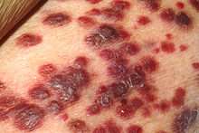
غرنة كابوزي - غرنة كابوزي
- حمى لاسا
- قرحة ليبشوتز (ulcus vulvae acutum)
- حصبة (rubeola، morbilli)
- Milker's nodule
- لقاح جدري الماء
- مليساء معدية
- ثؤلول راحي إخمصي
- هربس بسيط وليدي
- هربس نطاقي عيني
- أرف (contagious pustular dermatosis، ecthyma contagiosum، infectious labial dermatitis، sheep pox)
- أرف
- هربس الشفة (herpes labialis)
- Papular purpuric gloves and socks syndrome
- ثؤلول مصطبغ
- ألم عصبي تالي للهربس (zoster-associated pain)
- Post-vaccination follicular eruption
- وقس مترقي (vaccinia gangrenosum، vaccinia necrosum)
- Pseudocowpox
- ورام حليمي بالحنجرة (laryngeal papillomatosis)
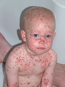
جدري الماء - حمى الوادي المتصدع
- طفح وردي (exanthem subitum، exanthema subitum، sixth disease)
- Roseola vaccinia
- حصبة ألمانية (German measles)
- حمى باباتاسية (Pappataci fever، phlebotomus fever)
- Sealpox
- جدري الماء (chickenpox)
- جدري (smallpox)
- ثؤلول مسطح (flat wart)
- ثؤلول إخمصي (plantar wart)
- ثؤلول (wart)
- Verrucae palmares et plantares
- Viral-associated trichodysplasia (ciclosporin-induced folliculodystrophy)
- متلازمة الهزال
- فيروس غرب النيل
- حماق (herpes zoster، shingles)
- هربس نطاقي


أمراض حزازانية[عدل]
الأمراض الحزازانية هي أمراض جلدية مرتبطة باضطرابات حزاز مسطح التهاببية شائعة فريدة، تصيب الجلد والأغشية المخاطية والأظافر والشعر.[71][72][73]
- حزاز مسطح حلقي
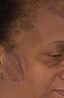
حزاز مسطح - حزاز مسطح ضموري
- حزاز مسطح (vesiculobullous lichen planus)
- حزاز مسطح
- Erythema dyschromicum perstans (ashy dermatosis، dermatosis cinecienta)
- التهاب الجلد الحزازاني ذو الخلايا العملاقة
- حزاز مسطح
- حزاز مسطح (lichen planus verrucosus)
- Idiopathic eruptive macular pigmentation
- حزاز مسطح
- حزاز مسطح (Nékam's disease)
- لطع الفرج
- Lichen nitidus
- حزاز مسطح (actinic lichen niditus، actinic lichen planus، lichen planus atrophicus annularis، lichen planus subtropicus، lichen planus tropicus، lichenoid melanodermatitis، lichenoid melanodermatosis، summertime actinic lichenoid eruption)
- حزاز مسطح
- حزاز مسطح
- حزاز مسطح
- Lichen ruber moniliformis
- حزاز متصلب (lichen sclerosus et atrophicus)
- حزاز مخطط (Blaschko linear acquired inflammatory skin eruption، linear lichenoid dermatosis)
- Lichen verrucosus et reticularis
- Lichenoid trikeratosis
- حزاز مسطح
- حزاز مسطح
- حزاز مسطح
- حزاز مسطح
- حزاز مسطح
- حزاز مسطح
- حزاز مسطح
- حزاز مسطح

أمراض لمفاوية[عدل]
Lymphoid-related cutaneous conditions are a group of disorders characterized by collections of خلية لمفاوية cells within the skin.[74]
- ابيضاض تائي الخلايا في البالغين
- Angiocentric lymphoma (extranodal natural killer cell lymphoma، nasal-type NK lymphoma، NK/T-cell lymphoma، polymorphic/malignant midline reticulosis)
- لموفوما الخلايا التائية ذات الأرومات المناعية الوعائية (angioimmunoblastic lymphadenopathy with dysproteinemia)
- لمفوما الخلايا القاتلة الطبيعية الأرومية
- لمفوما تائية الخلايا جلدية موجبة السي دي 30 (primary cutaneous anaplastic large cell lymphoma)
- Cutaneous lymphoid hyperplasia (borrelial lymphocytoma، lymphadenosis benigna cutis، lymphocytoma cutis، pseudolymphoma، pseudolymphoma of Spiegler and Fendt، sarcoidosis of Spiegler and Fendt، Spiegler–Fendt lymphoid hyperplasia، Spiegler–Fendt sarcoid)

Cutaneous lymphoid hyperplasia - Cutaneous lymphoid hyperplasia with bandlike and perivascular patterns
- Cutaneous lymphoid hyperplasia with nodular pattern (nodular pattern of cutaneous lymphoid hyperplasia)
- لمفوما الخلية البائية الكبيرة المنتشرة (primary cutaneous large B-cell lymphoma)
- ساركومة نقوية (chloroma، myeloid sarcoma)
- Granulomatous slack skin
- ابيضاض المشعر الخلاي
- لمفوما هودجكين
- سماك مكتسب (acquired ichthyosis)
- IgG4-related disease
- اللمفوما داخل الأوعية (angiotropic large cell lymphoma، intralymphatic lymphomatosis، intravascular lymphomatosis، malignant angioendotheliomatosis)
- Jessner lymphocytic infiltrate of the skin (benign lymphocytic infiltration of the skin، Jessner lymphocytic infiltration of the skin، Jessner–Kanof lymphocytic infiltration of the skin، lymphocytic infiltrate of Jessner)
- مرض كيكوتشي (histiocytic necrotizing lymphadenitis)
- Large plaque parapsoriasis (parapsoriasis en plaques)
- لمفوما لينيرت (lymphoepitheliod lymphoma)
- Leukemia cutis
- لمفوما الجلد
- ورم حبيبي لمفاوي
- كثرة الحطاطات اللمفومية
- كثرة المنسجات الخبيثة (histiocytic medullary reticulosis)
- لمفومات الخلايا البائية في المنطقة الهامشية
- لمفوما النسيج اللمفاوي المرتبط بالمخاطيات
- فطار فطراني
- لمفوما كبيرة الخلايا جلدية سالبة السي دي 30 لافطارية شبيهة بالفطر
- Nonspecific cutaneous conditions associated with leukemia (leukemid)
- Pagetoid reticulosis (acral mycoses fungoides، localized epidermotropic reticulosis، mycosis fungoides palmaris et plantaris، unilesional mycosis fungoides، Woringer–Kolopp disease)
- نخالية حزازانية مزمنة (chronic guttate parapsoriasis، chronic pityriasis lichenoides، dermatitis psoriasiformis nodularis، parapsoriasis chronica، parapsoriasis lichenoides chronica)
- نخالية حزازية وجدرية حادة (acute guttate parapsoriasis، acute parapsoriasis، acute pityriasis lichenoides، Mucha–Habermann disease، parapsoriasis acuta، parapsoriasis lichenoides et varioliformis acuta، parapsoriasis varioliformis)
- ورم البلازماويات
- Plasmacytosis
- Pleomorphic T-cell lymphoma (non-mycosis fungoides CD30− pleomorphic small/medium sized cutaneous T-cell lymphoma)
- كثرة الحمر الحقيقية (erythremia)
- Primary cutaneous follicular lymphoma (follicular center cell lymphoma، follicular center lymphoma)
- Primary cutaneous immunocytoma
- Primary cutaneous marginal zone lymphoma
- Retiform parapsoriasis
- لمفوما جلدية ثانوية ضخمة الخلايا موجبة السي دي 30
- مرض سيزاري
- مرض روزاي دورفمان (كثرة منسجات الجيوب الأنفية مع تضخم كبير للعقد اللمفية)
- Subcutaneous T-cell lymphoma (panniculitis-like T-cell lymphoma)
- Vesiculopustular eruption and leukemoid reaction in Down syndrome

وحمات وأمراض ميلانينية[عدل]
Melanocytic nevi and neoplasms are caused by either a proliferation of (1) خلية ميلانينيةs، or (2) خلية وحمية s، a form of melanocyte that lack dendritic processes.[75][76]
- Acral nevus (melanocytic nevus of acral skin، melanocytic nevus with intraepidermal ascent of cells)
- Amelanotic blue nevus (hypomelanotic blue nevus)
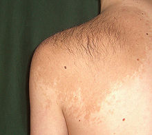
Becker's nevus - وحمة الخلية البالونية
- متلازمة بانيان- رايلي- روفالكابا
- Becker's nevus (Becker's melanosis، Becker's pigmentary hamartoma، nevoid melanosis، pigmented hairy epidermal nevus)
- Benign melanocytic nevus (banal nevus، common acquired melanocytic nevus، mole، nevocellular nevus، nevocytic nevus)
- Blue nevus (blue neuronevus، dermal melanocytoma، nevus bleu)
- Blue nevus of Jadassohn–Tièche (common blue nevus، nevus ceruleus)
- تلازمة كارني (LAMB syndrome، NAME syndrome)
- Cellular blue nevus
- Centrofacial lentiginosis
- شامة ميلانينية خلقية
- Deep penetrating nevus
- وحمة خلل التنسج (atypical mole، atypical nevus، B-K mole، Clark's nevus، dysplastic melanocytic nevus، nevus with architectural disorder)
- متلازمة وحمة خلل التنسج (B-K mole syndrome، familial atypical multiple mole–melanoma syndrome، familial melanoma syndrome)
- نمش (freckle)
- Epithelioid blue nevus
- Generalized lentiginosis
- Giant pigmented nevus (bathing trunk nevus، congenital nevomelanocytic nevus، garment nevus، giant hairy nevus، nevus pigmentosus et pilosus)
- وحمة هالية (leukoderma acquisitum centrifugum، perinevoid vitiligo، Sutton nevus)
- Hori's nevus (acquired bilateral nevus of Ota-like macules)
- Inherited patterned lentiginosis in black persons
- Ink spot lentigo (sunburn lentigo)
- Laugier–Hunziker syndrome
- Lentigo simplex (simple lentigo)
- Malignant blue nevus
- شامة ميلانينية خلقية
- تقران مثي
- Melanocytic tumors of uncertain malignant potential
- متلازمة نونان ذات التصبغات المتعددة
- Mucosal lentigines (labial and penile and vulvar melanosis، melanotic macules)
- وحمة إيتو (nevus fuscoceruleus acromiodeltoideus)
- وحمة أوتا (congenital melanosis bulbi، melanosis bulborum and aberrant dermal melanocytosis، nevus fuscoceruleus ophthalmomaxillaris، oculodermal melanocytosis، oculomucodermal melanocytosis)
- Nevus spilus (speckled lentiginous nevus، zosteriform lentiginous nevus)
- Partial unilateral lentiginosis (segmental lentiginosis)
- متلازمة بوتز-جيغرز
- Pigmented spindle cell nevus (pigmented spindle cell tumor of Reed، pigmented variant of Spitz nevus)
- Pseudomelanoma (recurrent melanocytic nevus، recurrent nevus)
- PUVA lentigines
- شامة ميلانينية خلقية
- وحمة سبيتز (benign juvenile melanoma، epithelioid and spindle cell nevus، Spitz's juvenile melanoma)
- بقعة كبدية (lentigo senilis، liver spot، old age spot، senile freckle)

الميلانوما[عدل]
ورم ميلانيني is a خباثة proliferation of melanocytes and the most aggressive type of skin cancer.[77][78][79]
- ورم ميلانيني نمشي طرفي
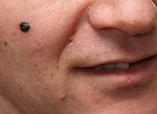
Nodular melanoma - Amelanotic melanoma
- Animal-type melanoma
- Desmoplastic melanoma (neurotropic melanoma، spindled melanoma)
- Lentigo maligna (lentiginous melanoma on sun-damaged skin)
- Lentigo maligna melanoma
- Melanoma with features of a Spitz nevus (Spitzoid melanoma)
- Melanoma with small nevus-like cells (small cell melanoma)
- Mucosal melanoma
- ورم ميلانيني وحماني
- Nodular melanoma
- Polypoid melanoma
- ورم ميلانيني
- Soft-tissue melanoma (clear-cell sarcoma، melanoma of the soft parts)
- ورم ميلانيني سطحي الانتشار (superficially spreading melanoma)
- ورم ميلانيني عنبي

أمراض متعلقة بالخلايا الوحيدة والخلايا الأكولة[عدل]
Monocyte- and macrophage-related cutaneous conditions are characterized علم الأنسجة by ارتشاح of the skin by خلية وحيدة and/or خلية أكولة كبيرة cells،[10] often divided into several categories، including ورم حبيبي،[80] كثرة المنسجات،[81] and غرناوية (ساركويد).[82]
- ورم حبيبي سافع (O'Brien granuloma)
- Annular elastolytic giant cell granuloma (giant cell elastophagocytosis، Meischer's granuloma، Miescher's granuloma of the face)
- ساركويد المحمرة للجلد
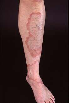
Generalized granuloma annulare - كثرة المنسجات الرأسية الحميدة (كثرة المنسجات مع أجسام مثل الدودة داخل السيتوبلازم).
- كثرة المنسجات الشبكية الخلقية ذاتية الشفاء (داء هاشيموتو-بريتزكر)
- ساركويد المحمرة للجلد
- ورم المنسجات الطفحي المتعمم
- Generalized granuloma annulare
- Giant cell reticulohistiocytoma (solitary reticulohistiocytoma، solitary reticulohistiocytosis)
- Granuloma annulare in HIV disease
- Granuloma multiforme (Mkar disease، granuloma multiforme (Leiker))
- داء هاند-شولر-كريستيان
- متلازمة هيرفوردت
- كثرة المنسجات المخاطية الوراثية المترقية
- ساركويد المحمرة للجلد
- ساركويد المحمرة للجلد
- كثرة المنسجات للخلايا غير المتمايزة
- Interstitial granulomatous drug reaction
- كثرة المنسجات لخلايا لانغر هانز(كثرة المنسجات إكس)
- داء ليترير سايوي
- Localized granuloma annulare
- متلازمة لوفغرين
- ذئبة تثليجية
- ساركويد المحمرة للجلد
- ساركويد المحمرة للجلد
- كثرة المنسجات الشبكية متعددة المراكز
- Necrobiotic xanthogranuloma (necrobiotic xanthogranuloma with paraproteinemia)
- كثرة منسجات غير إكسية
- ساركويد المحمرة للجلد
- الورم الأصفر الحطاطي
- Patch-type granuloma annulare (macular granuloma annulare)
- Perforating granuloma annulare
- كثرة المنسجات العقدية المترقية
- ورم كثرة المنسجات الشبكية
- ساركويد المحمرة للجلد (sarcoidosis in scars)
- كثرة منسجات البحر الأزرق
- Subcutaneous granuloma annulare (deep granuloma annulare، pseudorheumatoid nodule)
- ساركويد المحمرة للجلد (Darier–Roussy disease، Darier–Roussy sarcoid)
- غرناوية (ساركويد)
- ساركويد المحمرة للجلد
- الورم الأصفر المنتثر (متلازمة مونتغمري)

أمراض موسينية[عدل]
داء موسيني are a group of conditions caused by أدمة خلية ليفية يافعةs producing abnormally large amounts of غليكوز أمينوغليكان.[34]
- Acral persistent papular mucinosis
- Atypical lichen myxedematosus (intermediate lichen myxedematosus)
- Atypical tuberous myxedema (Jadassohn–Dosseker syndrome)
- Cutaneous focal mucinosis
- Cutaneous lupus mucinosis (papular and nodular mucinosis in lupus erythematosus، papular and nodular mucinosis of Gold، papulonodular mucinosis in lupus erythematosus)
- Discrete papular lichen myxedematosus
- داء موسيني ناتح
- Follicular mucinosis (alopecia mucinosa، mucinosis follicularis، Pinkus’ follicular mucinosis، Pinkus’ follicular mucinosis–benign primary form)
- Localized lichen myxedematosus
- كيسة مخاطانية (digital mucous cyst، mucous cyst)
- Myxoid lipoblastoma
- Neuropathia mucinosa cutanea
- Nodular lichen myxedematosus
- Papular mucinosis (generalized lichen myxedematosus، sclerodermoid lichen myxedematosus، scleromyxedema)
- Papular mucinosis of infancy (cutaneous mucinosis of infancy)
- داء موسيني محيط بالجريب
- Reticular erythematous mucinosis (midline mucinosis، plaque-like cutaneous mucinosis، REM syndrome)
- تصلب الجلد
- Self-healing juvenile cutaneous mucinosis
- Self-healing papular mucinosis
- Stiff skin syndrome (congenital fascial dystrophy)
أمراض عصبية جلدية[عدل]
Neurocutaneous conditions are due عضو جهاز عصبي disease or are طب نفسي in علم أسباب الأمراض.[83][84]
- ألم مزمن
- اضطراب التشوه الجسمي (dysmorphic syndrome، dysmorphophobia)
- Brachioradial pruritus
- المتلازمة التحويلية الشمية
- متلازمة الألم الناحي المركب (reflex sympathetic dystrophy)
- عدم الحساسية الخلقية للألم مع عدم التعرق
- داء طفيلي وهامي (delusions of parasitosis، Ekbom syndrome، monosymptomatic hypochondriacal psychosis)
- Dermatothlasia
- التهاب الجلد المفتعل (dermatitis artefacta، factitial dermatitis)

التهاب الجلد المفتعل - متلازمة الفم الحارق (burning mouth syndrome، burning tongue، orodynia)
- Levator ani syndrome
- قرحة عصبية تغذوية (neurotrophic ulcer، perforating ulcer of the foot)
- ألم الفخذ المذلي (Roth–Bernhardt disease)
- تسحج
- Notalgia paresthetica (hereditary localized pruritus، posterior pigmented pruritic patch، subscapular pruritus)
- Postencephalitic trophic ulcer
- Psychogenic pruritus
- خلل الوظائف المستقلة العائلي (familial dysautonomia)
- Scalp dysesthesia
- عصب وركي
- ألم الصفن
- تكهف النخاع (Morvan's disease)
- Traumatic neuroma (amputation neuroma)
- هوس نتف الشعر (trichotillosis)
- ألم عصب ثلاثي التوائم (tic douloureux)
- Trigeminal trophic lesion (trigeminal trophic syndrome)
- ألم الفرج (vestibulodynia)

أمراض غير معدية مرتبطة بنقص المناعة[عدل]
Noninfectious immunodeficiency-related cutaneous conditions are caused by خلية تائية and/or خلية بائية dysfunction.[85][86]
- متلازمة اللمفاويات العارية
- داء الورم الحبيبي المزمن (Bridges–Good syndrome، chronic granulomatous disorder، Quie syndrome)
- عوز مناعي شائع متغير (acquired hypogammaglobulinemia)
- عوز المتممة
- متلازمة دي جورج (DiGeorge anomaly، thymic hypoplasia)
- مرض الطعم ضد الثوي
- متلازمة غريسيلي
- Hyper-IgE syndrome (Buckley syndrome، Job syndrome)
- Immunodeficiency with hyper-IgM
- Immunodeficiency–centromeric instability–facial anomalies syndrome (ICF syndrome)
- عوز الغلوبولين المناعي إيه الانتقائي
- Isolated primary IgM deficiency
- Janus kinase 3 deficiency
- Leukocyte adhesion molecule deficiency
- LIG4 syndrome
- Myeloperoxidase deficiency
- Neutrophil immunodeficiency syndrome
- Nezelof syndrome (thymic dysplasia with normal immunoglobulins)
- متلازمة أومين
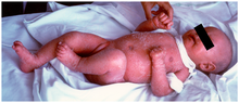
متلازمة أومين - Purine nucleoside phosphorylase deficiency
- عوز المناعة المشترك الشديد (alymphocytosis، Glanzmann–Riniker syndrome، severe mixed immunodeficiency syndrome، thymic alymphoplasia)
- متلازمة شواخمان دايموند
- Thymoma with immunodeficiency (Good syndrome)
- Transient hypogammaglobulinemia of infancy
- متلازمة وهيم (WHIM syndrome)
- متلازمة ويسكوت ألدريخ
- فقد غاماغلوبولين الدم المرتبط بالصبغي إكس (Bruton syndrome، sex-linked agammaglobulinemia)
- X-linked hyper-IgM syndrome
- فقد غاماغلوبولين الدم المرتبط بالصبغي إكس
- X-linked lymphoproliferative disease (Duncan's disease)
- قلة الخلايا المتعادلة

أمراض مرتبطة بنقص التغذية[عدل]
Nutrition-related cutaneous conditions are caused by سوء التغذية due to an improper or inadequate حمية غذائية.[87][88]
- Biotin deficiency
- كاروتينمية
- أحماض دهنية أساسية
- نقص الفوليك
- فرط الفيتامين أ
- نقص فيتامين أ (phrynoderma)
- عوز الحديد (طب)
- كواشيوركور
- وجود الليكوبين بالدم
- داء البول القيقبي
- سغل
- بلاغرا (pellagra، vitamin B3 deficiency)
- نقص السيلينيوم
- فيتامين ب1 (beriberi، thiamine deficiency)
- نقص فيتامين بي 12 (cyanocobalamin deficiency)
- فيتامين ب2 (ariboflavinosis، riboflavin deficiency)
- فيتامين ب6 (pyridoxine deficiency)
- فيتامين ب6 (pyridoxine excess)
- نقص فيتامين سي (scurvy)
- نقص فيتامين ك
- نقص الزنك
أمراض فرط التقرن الحطاطية الحرشفية[عدل]
اضطراب حطاطي حرشفي فرط التقرن cutaneous conditions are those that present with حطاطةs and مرض جلدي caused by a thickening of the طبقة متقرنة.[9]

- Confluent and reticulated papillomatosis (confluent and reticulated papillomatosis of Gougerot and Carteaud، familial cutaneous papillomatosis، familial occurrence of confluent and reticulated papillomatosis)
- Digitate dermatosis
- Drug-induced keratoderma
- احمرار الجلد (dermatitis exfoliativa، erythroderma، red man syndrome)
- ورام حليمي جلدي متورد
- Granular parakeratosis (axillary granular parakeratosis، intertriginous granular parakeratosis)
- Keratolysis exfoliativa (lamellar dyshidrosis، recurrent focal palmar peeling، recurrent palmar peeling)
- Keratosis punctata of the palmar creases (hyperkeratosis penetrans، hyperkeratosis punctata، keratodermia punctata، keratosis punctata، keratotic pits of the palmar creases، lenticular atrophia of the palmar creases، punctate keratosis of the palmar creases)
- حثل القرنية لميسمان
- Paraneoplastic keratoderma
- نخالية وردية (pityriasis rosea Gibert)
- نخالية شعرية حمراء (Devergie's disease، lichen ruber acuminatus، lichen ruber pilaris)
- Pure hair-nail type ectodermal dysplasia
- Small plaque parapsoriasis (chronic superficial dermatitis)
- راحة الكرش
- Xanthoerythrodermia perstans
تقرن جلد الراحة والأخمص المنتشر[عدل]
تقرن الجلد الراحي الأخمصي are a diverse group of hereditary and acquired تقرن الجلد in which there is فرط التقرن of the skin of the palms and soles.[89]
- Acrokeratoelastoidosis of Costa (keratoelastoidosis marginalis)
- تقرن الجلد الراحي الأخمصي (acquired aquagenic palmoplantar keratoderma، aquagenic syringeal acrokeratoderma، aquagenic wrinkling of the palms، transient reactive papulotranslucent acrokeratoderma)
- Bart–Pumphrey syndrome (palmoplantar keratoderma with knuckle pads and leukonychia and deafness)
- Camisa disease
- تقرن الجلد الراحي الأخمصي (striate palmoplantar keratoderma with woolly hair and cardiomyopathy، striate palmoplantar keratoderma with woolly hair and left ventricular dilated cardiomyopathy)
- Corneodermatoosseous syndrome (CDO syndrome)
- تقرن الجلد الراحي الأخمصي (palmoplantar keratoderma cum degeneratione granulosa Vörner، Vörner's epidermolytic palmoplantar keratoderma، Vörner keratoderma)
- تقرن الجلد الراحي الأخمصي (diffuse orthohyperkeratotic keratoderma، hereditary palmoplantar keratoderma، keratosis extremitatum progrediens، keratosis palmoplantaris diffusa circumscripta، tylosis، Unna–Thost disease، Unna–Thost keratoderma)
- Erythrokeratodermia variabilis (erythrokeratodermia figurata variabilis، keratosis extremitatum progrediens، keratosis palmoplantaris transgrediens et progrediens، Mendes da Costa syndrome، Mendes da Costa type erythrokeratodermia، progressive symmetric erythrokeratoderma)
- تقرن الجلد الراحي الأخمصي (acrokeratoelastoidosis lichenoides، degenerative collagenous plaques of the hand)
- Focal palmoplantar and gingival keratosis
- تقرن الجلد الراحي الأخمصي (focal epidermolytic palmoplantar keratoderma، hereditary painful callosities، hereditary painful callosity syndrome، keratosis follicularis، keratosis palmoplantaris nummularis، nummular epidermolytic palmoplantar keratoderma)
- متلازمة حاييم مونك (palmoplantar keratoderma with periodontitis and arachnodactyly and acro-osteolysis)
- Hidrotic ectodermal dysplasia (alopecia congenita with keratosis palmoplantaris، Clouston syndrome، Clouston's hidrotic ectodermal dysplasia، Fischer–Jacobsen–Clouston syndrome، keratosis palmaris with drumstick fingers، palmoplantar keratoderma and clubbing)
- متلازمة هاول إيفانز (familial keratoderma with carcinoma of the esophagus، focal non-epidermolytic palmoplantar keratoderma with carcinoma of the esophagus، palmoplantar ectodermal dysplasia type III، palmoplantar keratoderma associated with esophageal cancer، tylosis، tylosis–esophageal carcinoma)
- Hystrix-like ichthyosis–deafness syndrome (HID syndrome)
- Keratoderma climactericum (acquired plantar keratoderma، climacteric keratoderma، Haxthausen's disease)
- تقرن الجلد الراحي الأخمصي (Buschke–Fischer–Brauer disease، Davis Colley disease، keratoderma disseminatum palmaris et plantaris، keratosis papulosa، keratoderma punctatum، keratodermia punctata، keratoma hereditarium dissipatum palmare et plantare، palmar and plantar seed dermatoses، palmar keratoses، papulotranslucent acrokeratoderma، punctate keratoderma،punctate keratoses of the palms and soles، maculosa disseminata)
- Keratitis–ichthyosis–deafness syndrome (erythrokeratodermia progressiva Burns، ichthyosiform erythroderma with corneal involvement and deafness، KID syndrome)
- مرض ميليدا (acral keratoderma، Gamborg–Nielsen keratoderma، mutilating palmoplantar keratoderma of the Gamborg–Nielsen type، palmoplantar ectodermal dysplasia type VIII، palmoplantar keratoderma of the Norrbotten type)
- خلل تنسج البطين الأيمن المحدث لاضطراب النظم (diffuse non-epidermolytic palmoplantar keratoderma with woolly hair and cardiomyopathy، diffuse palmoplantar keratoderma with woolly hair and arrythmogenic right ventricular cardiomyopathy of Naxos، Naxos disease)
- تقرن الجلد الراحي الأخمصي (mutilating palmoplantar keratoderma with periorificial keratotic plaques، mutilating palmoplantar keratoderma with periorificial plaques، polykeratosis of Touraine)
- ثخن الأظفار الخلقي (Jadassohn–Lewandowsky syndrome)
- ثخن الأظفار الخلقي (Jackson–Lawler pachyonychia congenita، Jackson–Sertoli syndrome)
- تقرن الجلد الراحي الأخمصي (Charcot–Marie–Tooth disease with palmoplantar keratoderma and nail dystrophy)
- تقرن الجلد الراحي الأخمصي (Greither palmoplantar keratoderma، Greither syndrome، keratosis extremitatum hereditaria progrediens، keratosis palmoplantaris transgrediens et progrediens، Sybert keratoderma، transgrediens and progrediens palmoplantar keratoderma)
- متلازمة بابيلون لوفيفر (palmoplantar keratoderma with periodontitis)
- تقران منخرب
- تقرن الجلد الراحي الأخمصي
- Schöpf–Schulz–Passarge syndrome (eyelid cysts with palmoplantar keratoderma and hypodontia and hypotrichosis)
- تقرن الجلد الراحي الأخمصي (Huriez syndrome، palmoplantar keratoderma with scleroatrophy، palmoplantar keratoderma with sclerodactyly، scleroatrophic and keratotic dermatosis of the limbs، sclerotylosis)
- تقرن الجلد الراحي الأخمصي (acral keratoderma، Brünauer–Fuhs–Siemens type of palmoplantar keratoderma، focal non-epidermolytic palmoplantar keratoderma، keratosis palmoplantaris varians، palmoplantar keratoderma areata، palmoplantar keratoderma striata، Wachter keratoderma، Wachters palmoplantar keratoderma)
- تقرن الجلد الراحي الأخمصي (porokeratosis punctata palmaris et plantaris، punctate keratoderma، punctate porokeratosis of the palms and soles)
- Tyrosinemia type II (oculocutaneous tyrosinemia، Richner–Hanhart syndrome)
- متلازمة فوونكل (keratoderma hereditaria mutilans، keratoma hereditaria mutilans، mutilating keratoderma of Vohwinkel، mutilating palmoplantar keratoderma)
أمراض مرتبطة بالحمل[عدل]

Pregnancy-related cutaneous conditions are a group of skin changes observed during حمل.[90][91]
- قوباء هربسية الشكل
- ركود صفراوي داخل الكبد أثناء الحمل (cholestasis of pregnancy، jaundice of pregnancy، obstetric cholestasis، prurigo gravidarum)
- خط أسود
- شبيه الفقاع الحملي (gestational pemphigoid، herpes gestationis)
- حكاك الحمل (Besnier prurigo، early-onset prurigo of pregnancy، linear IgM dermatosis of pregnancy، papular dermatitis of pregnancy، prurigo of pregnancy، Spangler's papular dermatitis of pregnancy)
- التهاب الجريبات الحكي أثناء الحمل
- Pruritic urticarial papules and plaques of pregnancy (late-onset prurigo of pregnancy، polymorphic eruption of pregnancy، PUPPP syndrome، toxemic rash of pregnancy، toxic erythema of pregnancy)
- تجعدات صغيرة
الحكة[عدل]
حكة، commonly known as itchiness، is a sensation exclusive to the skin، and characteristic of many skin conditions.[92][93]
- Adult blaschkitis
- ألم مائي
- حكة مائية المنشأ
- حكة صفراوية
- Cholestatic pruritus
- حكة محدثة بالدواء
- Hydroxyethyl starch-induced pruritus
- التهاب الجلد العصبي (neurodermatitis)
- حكة بريونية
- حكاك عقيدي
- حكاك صباغي
- Prurigo simplex
- حكة شرجية
- حكة صفنية
- حكة فرجية
- نقاط حاكة (itchy points)
- حكة فروة الرأس
- حكة شيخوخية
- حكة يوريمية (renal pruritus)
الصدفية[عدل]
صداف is a common، chronic، and recurrent التهاب disease of the skin characterized by circumscribed، حمامى، dry، scaling مرض جلدي.[94][95][96]

- Annular pustular psoriasis
- صدفية
- Exanthematic pustular psoriasis
- صدفية بثرية معممة (pustular psoriasis of von Zumbusch)
- صدفية قطروية (eruptive psoriasis)
- صدفية الثنيات
- تقرن الجلد السيلاني (keratoderma blennorrhagicum)
- Localized pustular psoriasis
- صدفية حفاظية
- صدفية (chronic stationary psoriasis،plaque-like psoriasis)
- التهاب المفصل في الصدفية
- احمرار الجلد الصدفي (erythrodermic psoriasis)
- صدفية مثية (sebopsoriasis، seborrhiasis)
أمراض العدلات التفاعلية[عدل]
Reactive neutrophilic cutaneous conditions constitute a spectrum of disease mediated by خلية متعادلة، and typically associated with underlying diseases، such as داء الأمعاء الالتهابي and علم الدم خباثة.[97][98]

- حمامى عقدة
- Bowel-associated dermatosis–arthritis syndrome (bowel bypass syndrome، bowel bypass syndrome without bowel bypass، intestinal bypass arthritis–dermatitis syndrome)
- متلازمة مارشال
- Neutrophilic dermatosis of the dorsal hands (pustular vasculitis of the dorsal hands)
- Neutrophilic eccrine hidradenitis
- تقيح الجلد الغنغريني
- متلازمة بابا (PAPA syndrome)
- Rheumatoid neutrophilic dermatitis (rheumatoid neutrophilic dermatosis)
- Superficial granulomatous pyoderma
- الجلاد الحموي الحاد بالعدلات (acute febrile neutrophilic dermatosis)
- Sweet's syndrome-like dermatosis
- Vesicopustular dermatosis
أمراض راحة اليد والأخمص المستعصية[عدل]
Recalcitrant palmoplantar eruptions are skin conditions of the يد and أخمص (قدم) which are resistant to treatment.[34]
- التهاب الجلد الساعي (acrodermatitis continua، acrodermatitis continua of Hallopeau، acrodermatitis continua suppurativa Hallopeau، acrodermatitis perstans، dermatitis repens Crocker، Hallopeau's acrodermatitis، Hallopeau's acrodermatitis continua، pustular acrodermatitis)
- Infantile acropustulosis (acropustulosis of infancy)
- بثار الراحة والأخمص (persistent palmoplantar pustulosis، pustular psoriasis of the Barber type، pustular psoriasis of the extremities، pustulosis of palms and soles، pustulosis palmaris et plantaris)
- Pustular bacterid
أمراض ناتجة عن أخطاء استقلابية[عدل]
Skin conditions resulting from errors in metabolism are caused by إنزيم defects that lead to an accumulation or deficiency of various cellular components، including، but not limited to، حمض أمينيs، سكرياتs، and ليبيداتs.[16]
- برفريا متقطعة حادة
- سوء تغذية الكظرية وبيضاء الدماغ (Schilder's disease)
- بيلة ألكابتونية
- البرفيرية الحادة (Doss porphyria، plumboporphyria)
- B-mannosidase deficiency
- كاروتينمية
- متلازمة كاداسيل (CADASIL syndrome)
- داء الأورام الصفر المنتشرة الدماغية الوترية
- وجود السيترولين في الدم
- مرض غونتر (Gunther's disease)
- تفقع سكري المنشأ (bullosis diabeticorum، bullous eruption of diabetes mellitus)
- Diabetic cheiroarthropathy
- Diabetic dermopathy (shin spots)
- Dystrophic calcinosis cutis
- ورم أصفر
- بروتوبرفيرية مكونة للحمر

بروتوبرفيرية مكونة للحمر - مرض فابري (Anderson–Fabry disease، angiokeratoma corporis diffusum)
- عوز البروتين الشحمي العائلي (Tangier disease)
- اعتلال الأعصاب النشواني العائلي
- فرط شحميات الدم
- فرط شحميات الدم المركب (multiple-type hyperlipoproteinemia)
- Familial defective apolipoprotein B-100
- Familial dysbetalipoproteinemia (broad beta disease، remnant removal disease)
- Familial hypertriglyceridemia
- مرض فاربر (fibrocytic dysmucopolysaccharidosis، lipogranulomatosis)
- الداء الفوكوزيدي
- داء غوشيه
- نقرس (podagra، urate crystal arthropathy، urate deposition disease)
- مرض هارتناب (pellagra-like dermatosis)
- Hemodialysis-associated amyloidosis
- برفيرية كبدية مكونة للكريات الحمر
- الكوبروبورفيريا الوراثية
- Hereditary gelsolin amyloidosis
- Heredofamilial amyloidosis
- متلازمة هنتر
- متلازمة هيرلر (gargoylism، mucopolysaccharidosis type I)
- Hurler–Scheie syndrome (mucopolysaccharidosis type I H-S)
- نقص الهيالورونيداز (mucopolysaccharidosis type IX)
- Iatrogenic calcinosis cutis
- Idiopathic scrotal calcinosis (idiopathic calcified nodules of the scrotum)
- داء لافورا
- متلازمة لش-نيهان (juvenile gout)
- Lichen amyloidosis
- Limited joint mobility
- داء أورباخ فيته (hyalinosis cutis et mucosae، Urbach–Wiethe disease)
- نقص ليباز البروتين الدهني (chylomicronemia، chylomicronemia syndrome)
- Macular amyloidosis

ورم أصفر - متلازمة ماروتو لامي (mucopolysaccharidosis type VI)
- Medication-induced hyperlipoproteinemia
- Metastatic calcinosis cutis
- كلاس شبيه الدخينات
- متلازمة موركيو (mucopolysaccharidosis type IV)
- بلى حيوي شحماني (necrobiosis lipoidica diabeticorum)
- مرض نيمان-بيك
- Nodular amyloidosis
- ورم أصفر
- Normolipoproteinemic xanthomatosis
- Obstructive liver disease (xanthomatous biliary cirrhosis)
- تمغر
- Osteoma cutis
- ورم أصفر
- بيلة فينيولية
- Phytosterolemia (sitosterolemia)
- برفيرية جلدية آجلة
- Primary cutaneous amyloidosis
- Primary systemic amyloidosis
- Prolidase deficiency
- Pseudoporphyria (pseudoporphyria cutanea tarda)
- متلازمة سانفيليبو
- متلازمة شايا (mucopolysaccharidosis type I S)
- Secondary cutaneous amyloidosis
- Secondary systemic amyloidosis
- داء الإلعاب
- متلازمة سلاي (mucopolysaccharidosis type VII)
- Subepidermal calcified nodule (solitary congenital nodular calcification، Winer's nodular calcinosis)
- Transient erythroporphyria of infancy (purpuric phototherapy-induced eruption)

لويحة صفراء - Traumatic calcinosis cutis
- ورم أصفر (tuberous xanthoma)
- Tumoral calcinosis
- بورفيريا المبرقشة (mixed hepatic porphyria، mixed porphyria، South African genetic porphyria، South African porphyria)
- Verruciform xanthoma
- جلد شمعي
- داء ويلسون (hepatolenticular degeneration)
- لويحة صفراء (xanthelasma)
- Xanthoma diabeticorum
- ورم أصفر (plane xanthoma)
- Xanthoma striatum palmare
- ورم أصفر (tendinous xanthoma)
- ورم أصفر


أمراض ناتجة عن عوامل فيزياوية[عدل]
Skin conditions resulting from physical factors occur from a number of causes، including، but not limited to، hot and cold temperatures، احتكاك، and رطوبة.[34][99][100]
- جرح الحك
- زراق الأطراف
- حكاك سفعي (familial polymorphous light eruption of American Indians، hereditary polymorphous light eruption of American Indians، Hutchinson's summer prurigo، hydroa aestivale)
- حرق الهباء الجوي
- جلاد ضيائي عديد الأشكال
- Beryllium granuloma
- Black heel and palm (black heel، calcaneal petechiae، chromidrose plantaire، post-traumatic punctate intraepidermal hemorrhage، tache noir، talon noir)
- مسمار لحم (callosity، clavus، corn، heloma، heloma durum، heloma molle، intractable plantar keratosis، tyloma)
- صبغة الكربون

شرث - شرث (pernio، perniosis)
- التهاب الجلد الشعاعي المزمن (actinic reticuloid، chronic photosensitivity dermatitis، persistent light reactivity، photosensitive eczema)
- جاروسية
- نفطة غيبوبية
- قطع مرجاني
- نفطة متأخرة
- Dermatosis neglecta
- نفطة وذمة (edema bulla، hydrostatic bulla، stasis blister)
- حرق كهربائي
- Equestrian perniosis
- حمامى حرورية (fire stains، toasted skin syndrome)
- زراق الساقين الاحمراري
- متلازمة فافر-راكوشو (Favre–Racouchot disease، nodular cutaneous elastosis with cysts and comedones)
- ورم حبيبي للأجسام الغريبة
- Fracture blister
- نفطة الاحتكاك
- قضمة الصقيع
- Garrod's pad (violinist's viola pad)
- إصبع عازف القيثار
- Heel stick wound
- Heat edema
- حرق (إصابة)
- Hunan hand syndrome (chili burn)
- حصاف وقسي الشكل (Bazin's hydroa vacciniforme)
- Jogger's nipple
- جلاد ضيائي عديد الأشكال
- سرطان كايرو
- سرطان كانج
- قرحة كانجري
- إصابة الصاعقة
- Loop mark
- Magnetic resonance imaging burn (MRI burn)
- ورم حبيبي زئبقي
- جاروسية (miliaria crystalline، sudamina)
- جاروسية (mammillaria)
- جاروسية
- جاروسية (heat rash، prickly heat)
- Narcotic dermopathy
- جاروسية
- Painful fat herniation (painful piezogenic pedal papules، piezogenic papules)
- سرطان وقود الخث
- تشيخ ضوئي (dermatoheliosis)
- تحسس ضوئي مع عدوى فيروس العوز المناعي البشري
- التهاب الجلد القطراني الضوئي السمي
- التهاب الجلد الضوئي النباتي (Berloque dermatitis)
- Pinch mark
- جلاد ضيائي عديد الأشكال (polymorphic light eruption)
- جاروسية
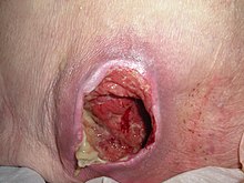
قرحة فراش - Postoperative hematoma
- قرحة فراش (decubitus ulcer)
- شواك كاذب أسود
- Pseudoverrucous papules and nodules
- Pulling boat hands
- PUVA-induced acrobullous dermatosis
- Runner's rump
- Sclerosing lymphangiitis
- Silica granuloma
- Silicone granuloma
- Skin pop scar
- Skin track
- رضة
- حمامى شمسية
- وشم السخام
- نفاخ تحت الجلد
- نفطة مص
- حرق شمسي
- Surfer's knots
- وشم
- ورم دموي تحت الظفر
- حرق حراري
- Traumatic asphyxia
- قدم الخندق
- جاروسية
- Tropical immersion foot (paddy foot، paddy-field foot)
- Turf toe
- جلاد اليورانيوم
- UV-sensitive syndrome
- Vibration white finger (dead finger، hand–arm vibration syndrome)
- Warm water immersion foot
- Weathering nodule of ear
- أذن قرنبيطية (cauliflower ear، traumatic auricular hematoma)
- Zirconium granuloma


أمراض محفزة بإشعاع مؤين[عدل]
Ionizing radiation-induced cutaneous conditions result from exposure to إشعاع مؤين.[101]
- حرق إشعاعي
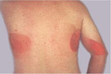
كشف فلوري - سرطان ناجم عن الإشعاع
- حرق إشعاعي
- حرق إشعاعي
- كشف فلوري
- حرق إشعاعي
- سرطان ناجم عن الإشعاع
- حرق إشعاعي (radiodermatitis)
- حرق إشعاعي
- حرق إشعاعي
- ندبة ضخامية
- جدرة
- قشيعة

الشرى والوذمة الوعائية[عدل]
شرى is a vascular reaction of the skin characterized by the appearance of مرض جلدي، which are firm، elevated swelling of the skin.[102] وذمة وعائية، which can occur alone or with urticaria، is characterized by a well-defined، edematous swelling that involves subcutaneous tissues، abdominal organs، and/or upper airway.[103]
- Acquired C1 esterase inhibitor deficiency
- شرى
- Adrenergic urticaria
- صدمة الحساسية
- الشرية المائية
- شرى كوليني
- شرى (ordinary urticaria)
- شرى البرد
- كتوبية الجلد (dermographism)
- Episodic angioedema with eosinophilia (Gleich's syndrome)
- شرى (exercise-induced urticaria)
- شرى غلفانية
- الشرى الحراري
- وذمة وعائية وراثية (Quincke's edema)
- Localized heat contact urticaria
- شرى
- شري فيزيائي
- شرى البرد
- Pressure urticaria (delayed pressure urticaria)
- شرى البرد
- متلازمة شنيتزلر
- شرى البرد
- شرى شمسي
- متلازمة تسرب شعيري
- Urticarial allergic eruption
- Urticaria-like follicular mucinosis
- Vibratory angioedema
أمراض مرتبطة بالأوعية الدموية[عدل]
Vascular-related cutaneous conditions result from dysfunction of the دم and/or وعاء دمويs in the dermis، or وعاء لمفي in the نسيج تحت الجلدs.[9][104][105]
- متلازمة أجيناس
- Acroangiodermatitis (acroangiodermatitis of Mali، Mali acroangiodermatitis، Pseudo-Kaposi's sarcoma)
- وذمة نزفية حادة في الرضع (acute hemorrhagic edema of childhood، Finkelstein's disease، infantile postinfectious iris-like purpura and edema، medallion-like purpura، purpura en cocarde avec oedema، Seidlmayer syndrome)
- قرحة القصور الشرياني (ischemic ulcer)
- Arteriosclerosis obliterans
- بقع بيير
- Blueberry muffin baby
- متلازمة ويبرن مايسون (Wyburn–Mason syndrome)
- Bullous lymphedema
- التهاب وعائي فقاعي بالأوعية الصغيرة (bullous variant of small vessel vasculitis)
- التأق التكلسي
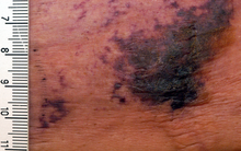
التأق التكلسي - الحدبة المصلية الدموية
- صمة كولسترولية (warfarin blue toe syndrome)
- Cobb syndrome
- Corona phlebectatica
- Cryofibrinogenemic purpura
- Cryoglobulinemic purpura
- Cryoglobulinemic vasculitis
- التهاب الأوعية الصغيرة في الجلد (cutaneous leukocytoclastic angiitis، cutaneous leukocytoclastic vasculitis، cutaneous necrotizing venulitis، hypersensitivity angiitis)
- خثار وريدي عميق
- تخثر منتشر داخل الأوعية
- Doucas and Kapetanakis pigmented purpura
- Drug-induced purpura
- Drug-induced thrombocytopenic purpura
- Eczematid-like purpura of Doucas and Kapetanakis
- خزب وبائي
- Erythema elevatum diutinum
- احمرار الأطراف المؤلم (acromelalgia، erythermalgia)
- Factitial lymphedema (hysterical edema)
- Fibrinolysis syndrome (defibrinating syndrome، hypofibrinogenemia)
- فرفرية
- Generalized essential telangiectasia (general essential telangiectasia)
- التهاب الشريان ذو الخلايا العملاقة
- Gougerot–Blum syndrome (pigmented purpuric lichenoid dermatitis، pigmented purpuric lichenoid dermatitis of Gougerot and Blum)
- Harlequin color change
- قرحة مكونة للدم
- Hennekam syndrome (Hennekam lymphangiectasia-lymphedema syndrome، intestinal lymphagiectasia-lymphedema-mental retardation syndrome)
- طفح جلدي نزفي (anaphylactoid purpura، purpura rheumatica، Schönlein–Henoch purpura)

طفح جلدي نزفي - توسع الشعيرات النزفي الوراثي (Osler's disease، Osler–Weber–Rendu disease)
- فرفرية قليلة الصفيحات مجهولة السبب (autoimmune thrombocytopenic purpura، Werlhof's disease)
- فرفرية هينوخ-شونلاين
- داء كاواساكي (mucocutaneous lymph node syndrome)
- Levamisole-induced vasculitis
- Lichen aureus (lichen purpuricus)
- تزرق عنقودي
- تزرق شبكي
- Livedoid dermatitis (embolia cutis medicamentosa، Nicolau syndrome)
- التهاب وعائي تزرقي (atrophie blanche، livedo reticularis with summer ulceration، livedoid vasculitis، PURPLE syndrome، segmental hyalinizing vasculitis)
- Lymphedema praecox
- متلازمة وذمة ازدواجية الأهداب اللمفية
- متلازمة مافوتشي
- Majocchi's disease (purpura annularis telangiectodes، purpura annularis telangiectodes of Majocchi)
- كثرة الحطاطات الضمورية الخبيثة (Degos' disease)
- متلازمة مارشال وايت
- Meige lymphedema
- التهاب الأوعية المجهري (microscopic polyarteritis، microscopic polyarteritis nodosa)
- داء موندور (Mondor's syndrome of superficial thrombophlebitis)
- قرحة القدم السكرية (mal perforans)
- Njolstad syndrome
- مرض ميلروي (hereditary lymphedema، Milroy disease)
- Obstructive purpura
- فرفرية انتصابية (stasis purpura)
- متلازمة التكدم المؤلم (autoerythrocyte sensitization، Gardner–Diamond syndrome، psychogenic purpura)
- متلازمة باركس ويبر
- Paroxysmal hand hematoma (Achenbach syndrome)
- بيلة هيمغلوبينية انتيابية ليلية
- التهاب الشرايين العقدي المتعدد (panarteritis nodosa، periarteritis nodosa)
- المتلازمة التالية لبضع التأمور
- Perinatal gangrene of the buttock
- Pigmentary purpuric eruptions (progressive pigmentary dermatosis، progressive pigmenting purpura، purpura pigmentosa chronica)
- Postinflammatory lymphedema
- متلازمة ستيوارت–تريفيز (Stewart–Treves syndrome)
- فرفرية خاطفة (purpura gangrenosa)
- فرفرية
- Purpuric agave dermatitis
ظاهرة رينو - ظاهرة رينو
- ظاهرة رينو (primary Raynaud's phenomenon)
- Reactive angioendotheliomatosis
- داء شامبرغ (progressive pigmentary dermatosis of Schamberg، purpura pigmentosa progressiva، Schamberg's purpura)
- Secondary lymphedema
- Septic thrombophlebitis
- Sinusoidal hemangioma
- متلازمة سنيدون (idiopathic livedo reticularis with cerebrovascular accidents)
- Solar purpura (actinic purpura، senile purpura)
- التهاب الجلد الركودي (congestion eczema، gravitational dermatitis، gravitational eczema، stasis eczema، varicose eczema)
- التهاب وريد خثاري سطحي
- التهاب الشرايين تاكاياسو (aortic arch syndrome، pulseless disease)
- التهاب الشريان ذو الخلايا العملاقة (cranial arteritis، Horton's disease)
- مرض بورغر (Buerger's disease)
- فرفرية نقص الصفيحات التخثرية (Moschcowitz syndrome)
- Traumatic purpura
- علامة تروسو للخباثة
- Unilateral nevoid telangiectasia (nevoid telangiectasia)
- التهاب الأوعية الدموية الشروي (chronic urticaria as a manifestation of venulitis، hypocomplementemic urticarial vasculitis syndrome، hypocomplementemic vasculitis، unusual lupus-like syndrome)
- قرحة وريدية
- Waldenström hyperglobulinemic purpura (purpura hyperglobulinemica)
- وجود الغلوبيولين الكبروي في الدم المنسوب لفالدنشتروم
- ورام حبيبي ويغنري
- متلازمة الأظافر الصفراء (primary lymphedema associated with yellow nails and pleural effusion)


ملاحظات[عدل]
- ^ Any given cutaneous condition is only included once within this list.
- ^ قوس are used to indicate other names by which a condition is known. If there are multiple alternative names for a condition, they are separated by commas within the parenthetical.
- ^ Citations for any given condition name and/or alternative name(s) may be found within the condition's respective article.
- ^ This list uses American English; therefore, the symbols æ and œ, which are common to British English, are not used, but, rather, simplified to a single E. For example, the spelling of وحمة is favored over وحمة، استسقاء (طب) over استسقاء (طب), and so forth. For more information, see American and British English differences.
- ^ لغة إنجليزية names are included within this list when those terms are found in English دليل طبي. Inclusion of حب الشباب العصبي السحجي (لغة فرنسية), داء عليقي (لغة ألمانية), and داء عليقي (لغة ملايو) represent examples of this عرف (مفهوم).
- ^ Abbreviations for condition names commonly described in medical literature with an اسم تاجي are included within this list.
- ^ Within this list, the term جسم مضاد is abbreviated to جسم مضاد when used as a prefix to a specific جسم مضاد (i.e. غلوبيولين مناعي A، جسم مضاد، كريين مناعي هـ، كريين مناعي ج, and كريين مناعي م).
- ^ Within this list, the terms human immunodeficiency virus and acquired immunodeficiency syndrome are abbreviated to فيروس العوز المناعي البشري and إيدز, respectively.
طالع أيضا[عدل]
المصادر[عدل]
- ^ أ ب ت ث ج Miller, Jeffrey H.; Marks, James G. (2006). Lookingbill and Marks' Principles of Dermatology. Saunders. ISBN:1-4160-3185-5.
{{استشهاد بكتاب}}: صيانة الاستشهاد: أسماء متعددة: قائمة المؤلفين (link) - ^ Lippens S, Hoste E, Vandenabeele P, Agostinis P, Declercq W؛ Hoste؛ Vandenabeele؛ Agostinis؛ Declercq (أبريل 2009). "Cell death in the skin". Apoptosis. ج. 14 ع. 4: 549–69. DOI:10.1007/s10495-009-0324-z. PMID:19221876.
{{استشهاد بدورية محكمة}}: صيانة الاستشهاد: أسماء متعددة: قائمة المؤلفين (link) - ^ أ ب ت ث Burns, Tony; et al. (2006) Rook's Textbook of Dermatology CD-ROM. Wiley-Blackwell. ISBN 1-4051-3130-6.
- ^ Paus R, Cotsarelis G؛ Cotsarelis (1999). "The biology of hair follicles". N Engl J Med. ج. 341 ع. 7: 491–7. DOI:10.1056/NEJM199908123410706. PMID:10441606.
- ^ Goldsmith, Lowell A. (1983). Biochemistry and physiology of the skin. Oxford University Press. ISBN:0-19-261253-0.
- ^ Fuchs E (فبراير 2007). "Scratching the surface of skin development". Nature. ج. 445 ع. 7130: 834–42. Bibcode:2007Natur.445..834F. DOI:10.1038/nature05659. PMC:2405926. PMID:17314969.
- ^ Fuchs E, Horsley V؛ Horsley (أبريل 2008). "More than one way to skin ". Genes Dev. ج. 22 ع. 8: 976–85. DOI:10.1101/gad.1645908. PMC:2732395. PMID:18413712.
- ^ أ ب ت ث ج Freedberg, Irwin M.;؛ وآخرون (2003). Fitzpatrick's Dermatology in General Medicine. McGraw-Hill. ISBN:0-07-138076-0.
{{استشهاد بكتاب}}: صيانة الاستشهاد: أسماء متعددة: قائمة المؤلفين (link) صيانة الاستشهاد: علامات ترقيم زائدة (link) - ^ أ ب ت ث Bolognia, Jean L.;؛ وآخرون (2007). Dermatology. St. Louis: Mosby. ISBN:1-4160-2999-0.
{{استشهاد بكتاب}}: صيانة الاستشهاد: أسماء متعددة: قائمة المؤلفين (link) صيانة الاستشهاد: علامات ترقيم زائدة (link) - ^ أ ب ت ث Rapini, Ronald P. (2005). Practical dermatopathology. Elsevier Mosby. ISBN:0-323-01198-5. مؤرشف من الأصل في 2022-08-18.
- ^ Grant-Kels JM (2007). Color Atlas of Dermatopathology (Dermatology: Clinical & Basic Science). Informa Healthcare. ص. 163. ISBN:0-8493-3794-1.
- ^ Ryan، T (1991). "Cutaneous Circulation". في Goldsmith، Lowell A (المحرر). Physiology, biochemistry, and molecular biology of the skin (ط. 2nd). New York: Oxford University Press. ص. 1019. ISBN:0-19-505612-4.
- ^ Swerlick RA, Lawley TJ؛ Lawley (يناير 1993). "Role of microvascular endothelial cells in inflammation". J. Invest. Dermatol. ج. 100 ع. 1: 111S–115S. DOI:10.1038/jid.1993.33. PMID:8423379.
- ^ أ ب ت ث Lynch, Peter J. (1994). Dermatology. Williams & Wilkins. ISBN:0-683-05252-7.
- ^ King, L.S. (1954). "What Is Disease?". Philosophy of Science. ج. 21 ع. 3: 193–203. DOI:10.1086/287343.
- ^ أ ب Bluefarb, Samuel M. (1984). Dermatology. Upjohn Co. ISBN:0-89501-004-6.
- ^ Tilles G, Wallach D; Wallach (1989). "[The history of nosology in dermatology]". Ann Dermatol Venereol (بالفرنسية). 116 (1): 9–26. PMID:2653160.
- ^ Lambert WC, Everett MA؛ Everett (أكتوبر 1981). "The nosology of parapsoriasis". J. Am. Acad. Dermatol. ج. 5 ع. 4: 373–95. DOI:10.1016/S0190-9622(81)70100-2. PMID:7026622.
- ^ Jackson R (1977). "Historical outline of attempts to classify skin diseases". Can Med Assoc J. ج. 116 ع. 10: 1165–8. PMC:1879511. PMID:324589.
- ^ Copeman PW (فبراير 1995). "The creation of global dermatology". J R Soc Med. ج. 88 ع. 2: 78–84. PMC:1295100. PMID:7769599.
- ^ Fitzpatrick, Thomas B.; Klauss Wolff; Wolff, Klaus Dieter; Johnson, Richard R.; Suurmond, Dick; Richard Suurmond (2005). Fitzpatrick's color atlas and synopsis of clinical dermatology. McGraw-Hill Medical Pub. Division. ISBN:0-07-144019-4.
{{استشهاد بكتاب}}: صيانة الاستشهاد: أسماء متعددة: قائمة المؤلفين (link) - ^ Werner B (Aug 2009). "[Skin biopsy and its histopathologic analysis: Why? What for? How? Part I]". An Bras Dermatol (بالبرتغالية). 84 (4): 391–5. PMID:19851671.
- ^ Werner B (Oct 2009). "[Skin biopsy with histopathologic analysis: why? what for? how? part II]". An Bras Dermatol (بالبرتغالية). 84 (5): 507–13. DOI:10.1590/S0365-05962009000500010. PMID:20098854.
- ^ Xiaowei Xu; Elder, David A.; Rosalie Elenitsas; Johnson, Bernett L.; Murphy, George E. (2008). Lever's Histopathology of the Skin. Hagerstwon, MD: Lippincott Williams & Wilkins. ISBN:0-7817-7363-6.
{{استشهاد بكتاب}}: صيانة الاستشهاد: أسماء متعددة: قائمة المؤلفين (link) - ^ Weedon's Skin Pathology, 2-Volume Set: Expert Consult - Online and Print. Edinburgh: Churchill Livingstone. 2009. ISBN:0-7020-3941-1.
- ^ أ ب ت ث ج David J. DiCaudo; Dirk Elston MD; Dirk M. Elston; Tammie Ferringer; Christine J. Ko; Christine Ko MD; Steven Peckham; Whitney A. High (2009). Dermatopathology. Philadelphia: Saunders. ISBN:0-7020-3023-6.
{{استشهاد بكتاب}}: صيانة الاستشهاد: أسماء متعددة: قائمة المؤلفين (link) - ^ Rustin MH (1990). "Dermatology". Postgrad Med J. ج. 66 ع. 781: 894–905. DOI:10.1136/pgmj.66.781.894. PMC:2429766. PMID:2148371.
- ^ Feldman S, Careccia RE, Barham KL, Hancox J؛ Careccia؛ Barham؛ Hancox (2004). "Diagnosis and treatment of acne". Am Fam Physician. ج. 69 ع. 9: 2123–30. PMID:15152959.
{{استشهاد بدورية محكمة}}: صيانة الاستشهاد: أسماء متعددة: قائمة المؤلفين (link) - ^ Fietta P (2004). "Autoinflammatory diseases: the hereditary periodic fever syndromes". Acta Biomed. ج. 75 ع. 2: 92–9. PMID:15481697.
- ^ Centola M, Aksentijevich I, Kastner DL؛ Aksentijevich؛ Kastner (1998). "The hereditary periodic fever syndromes: molecular analysis of a new family of inflammatory diseases". Hum Mol Genet. ج. 7 ع. 10: 1581–8. DOI:10.1093/hmg/7.10.1581. PMID:9735379. مؤرشف من الأصل في 2010-09-06.
{{استشهاد بدورية محكمة}}: صيانة الاستشهاد: أسماء متعددة: قائمة المؤلفين (link) - ^ Chan، Lawrence S. (30 مارس 2009). Blistering Skin Diseases (ط. 1). Manson Publishing Ltd. ISBN:978-1-84076-066-8. مؤرشف من الأصل في 2012-11-11.
- ^ Yeh SW, Ahmed B, Sami N, Razzaque Ahmed A؛ Ahmed؛ Sami؛ Razzaque Ahmed (2003). "Blistering disorders: diagnosis and treatment". Dermatol Ther. ج. 16 ع. 3: 214–23. DOI:10.1046/j.1529-8019.2003.01631.x. PMID:14510878.
{{استشهاد بدورية محكمة}}: صيانة الاستشهاد: أسماء متعددة: قائمة المؤلفين (link) - ^ Eming R, Hertl M؛ Hertl؛ Autoimmune Diagnostics Working Group (2006). "Autoimmune bullous disorders". Clin Chem Lab Med. ج. 44 ع. 2: 144–9. DOI:10.1515/CCLM.2006.027. PMID:16475898.
- ^ أ ب ت ث James, William D.؛ وآخرون (2006). Andrews' Diseases of the Skin: Clinical Dermatology. Saunders Elsevier. ISBN:0-7216-2921-0.
- ^ أ ب ت ث ج Christine J. Ko; Schwarzenberger, Kathryn; Werchniak, Andrew E. (2009). General dermatology. Philadelphia: Saunders. ISBN:0-7020-3093-7.
{{استشهاد بكتاب}}: صيانة الاستشهاد: أسماء متعددة: قائمة المؤلفين (link) - ^ Aronson IK, Tharp MD؛ Worobec (2010). "Diagnosis and Treatment of Panniculitis". Dermatologic Therapy. ج. 23 ع. 4: 317–434. DOI:10.1111/j.1529-8019.2010.01331.x. ISSN:1529-8019.
- ^ Requena L, Yus ES؛ Yus (2001). "Panniculitis. Part I. Mostly septal panniculitis". J Am Acad Dermatol. ج. 45 ع. 2: 163–83, quiz 184–6. DOI:10.1067/mjd.2001.114736. PMID:11464178.
- ^ Requena L, Sánchez Yus E؛ Sánchez Yus (2001). "Panniculitis. Part II. Mostly lobular panniculitis". J Am Acad Dermatol. ج. 45 ع. 3: 325–61, quiz 362–4. DOI:10.1067/mjd.2001.114735. PMID:11511831.
- ^ Phelps RG, Shoji T؛ Shoji (2001). "Update on panniculitis". Mt Sinai J Med. ج. 68 ع. 4–5: 262–7. PMID:11514913. مؤرشف من الأصل في 2009-02-14.
- ^ Küster W, Traupe H; Traupe (Sep 1988). "[Clinical aspects and genetics of congenital skin defects]". Hautarzt (بالألمانية). 39 (9): 553–63. PMID:3053531.
- ^ Andrea L. Zaenglein; MD, Howard Gimbel; Albert C. Yan (2008). Pediatric Dermatology: Requisites in Dermatology. Saunders Ltd. ISBN:0-7020-3022-8.
{{استشهاد بكتاب}}: صيانة الاستشهاد: أسماء متعددة: قائمة المؤلفين (link) - ^ Crofford, Leslie J.; Klippel, John H.; Weyand, Cornelia M.; Stone, John F. (2001). Primer on the rheumatic diseases. Atlanta, GA: Arthritis Foundation. ISBN:0-912423-29-3.
{{استشهاد بكتاب}}: صيانة الاستشهاد: أسماء متعددة: قائمة المؤلفين (link) - ^ Ushiki T (2002). "Collagen fibers, reticular fibers and elastic fibers. A comprehensive understanding from a morphological viewpoint". Arch Histol Cytol. ج. 65 ع. 2: 109–26. DOI:10.1679/aohc.65.109. PMID:12164335. مؤرشف من الأصل (PDF) في 2019-06-20.
- ^ Alsaad KO, Ghazarian D؛ Ghazarian (2005). "My approach to superficial inflammatory dermatoses". J Clin Pathol. ج. 58 ع. 12: 1233–41. DOI:10.1136/jcp.2005.027151. PMC:1770784. PMID:16311340.
- ^ Wüthrich B, Cozzio A, Roll A, Senti G, Kündig T, Schmid-Grendelmeier P؛ Cozzio؛ Roll؛ Senti؛ Kündig؛ Schmid-Grendelmeier (2007). "Atopic eczema: genetics or environment?". Ann Agric Environ Med. ج. 14 ع. 2: 195–201. PMID:18247450.
{{استشهاد بدورية محكمة}}: صيانة الاستشهاد: أسماء متعددة: قائمة المؤلفين (link) - ^ Roosterman D, Goerge T, Schneider SW, Bunnett NW, Steinhoff M؛ Goerge؛ Schneider؛ Bunnett؛ Steinhoff (2006). "Neuronal control of skin function: the skin as a neuroimmunoendocrine organ". Physiol Rev. ج. 86 ع. 4: 1309–79. DOI:10.1152/physrev.00026.2005. PMID:17015491.
{{استشهاد بدورية محكمة}}: صيانة الاستشهاد: أسماء متعددة: قائمة المؤلفين (link) - ^ Spergel JM (أغسطس 2010). "From atopic dermatitis to asthma: the atopic march". Ann. Allergy Asthma Immunol. ج. 105 ع. 2: 99–106, quiz 107–9, 117. DOI:10.1016/j.anai.2009.10.002. PMID:20674819. مؤرشف من الأصل في 2019-12-16.
- ^ Saint-Mezard P, Rosieres A, Krasteva M؛ وآخرون (2004). "Allergic contact dermatitis". Eur J Dermatol. ج. 14 ع. 5: 284–95. PMID:15358566. مؤرشف من الأصل في 2020-03-16.
{{استشهاد بدورية محكمة}}: صيانة الاستشهاد: أسماء متعددة: قائمة المؤلفين (link) - ^ Krasteva M, Kehren J, Sayag M؛ وآخرون (1999). "Contact dermatitis II. Clinical aspects and diagnosis". Eur J Dermatol. ج. 9 ع. 2: 144–59. PMID:10066966. مؤرشف من الأصل في 2020-03-16.
{{استشهاد بدورية محكمة}}: صيانة الاستشهاد: أسماء متعددة: قائمة المؤلفين (link) - ^ Sharma VK, Asati DP؛ Asati (2010). "Pediatric contact dermatitis". Indian J Dermatol Venereol Leprol. ج. 76 ع. 5: 514–20. DOI:10.4103/0378-6323.69070. PMID:20826990.
{{استشهاد بدورية محكمة}}: صيانة الاستشهاد: دوي مجاني غير معلم (link) - ^ Buxton PK (1987). "ABC of dermatology. Eczema and dermatitis". Br Med J (Clin Res Ed). ج. 295 ع. 6605: 1048–51. DOI:10.1136/bmj.295.6605.1048. PMC:1248082. PMID:3120868.
- ^ Mengesha YM, Bennett ML؛ Bennett (2002). "Pustular skin disorders: diagnosis and treatment". Am J Clin Dermatol. ج. 3 ع. 6: 389–400. DOI:10.2165/00128071-200203060-00003. PMID:12113648.
- ^ Schwartz RA, Janusz CA, Janniger CK؛ Janusz؛ Janniger (2006). "Seborrheic dermatitis: an overview". Am Fam Physician. ج. 74 ع. 1: 125–30. PMID:16848386.
{{استشهاد بدورية محكمة}}: صيانة الاستشهاد: أسماء متعددة: قائمة المؤلفين (link) - ^ Costin GE, Hearing VJ؛ Hearing (2007). "Human skin pigmentation: melanocytes modulate skin color in response to stress". FASEB J. ج. 21 ع. 4: 976–94. DOI:10.1096/fj.06-6649rev. PMID:17242160.
- ^ Yamaguchi Y, Brenner M, Hearing VJ؛ Brenner؛ Hearing (2007). "The regulation of skin pigmentation". J Biol Chem. ج. 282 ع. 38: 27557–61. DOI:10.1074/jbc.R700026200. PMID:17635904.
{{استشهاد بدورية محكمة}}: صيانة الاستشهاد: أسماء متعددة: قائمة المؤلفين (link) - ^ Davis EC, Callender VD؛ Callender (يوليو 2010). "Postinflammatory hyperpigmentation: a review of the epidemiology, clinical features, and treatment options in skin of color". J Clin Aesthet Dermatol. ج. 3 ع. 7: 20–31. PMC:2921758. PMID:20725554.
- ^ Goolamali SK (1985). "Drug eruptions". Postgrad Med J. ج. 61 ع. 720: 925–33. DOI:10.1136/pgmj.61.720.925. PMC:2418295. PMID:2932688.
- ^ Svensson CK, Cowen EW, Gaspari AA؛ Cowen؛ Gaspari (2001). "Cutaneous drug reactions". Pharmacol Rev. ج. 53 ع. 3: 357–79. PMID:11546834. مؤرشف من الأصل في 2009-10-13.
{{استشهاد بدورية محكمة}}: صيانة الاستشهاد: أسماء متعددة: قائمة المؤلفين (link) - ^ Ramdial PK, Naidoo DK؛ Naidoo (يونيو 2009). "Drug-induced cutaneous pathology". J. Clin. Pathol. ج. 62 ع. 6: 493–504. DOI:10.1136/jcp.2008.058289. PMID:19155238.
- ^ Reichrath J (يوليو 2009). "The skin is a fascinating endocrine organ". Dermatoendocrinol. ج. 1 ع. 4: 195–6. DOI:10.4161/derm.1.4.9653. PMC:2835874. PMID:20592790.
- ^ Slominski A, Wortsman J؛ Wortsman (2000). "Neuroendocrinology of the skin". Endocr Rev. ج. 21 ع. 5: 457–87. DOI:10.1210/er.21.5.457. PMID:11041445. مؤرشف من الأصل في 2011-04-30.
- ^ Heymann WR (يوليو 2006). "Eosinophilic dermatoses". J. Am. Acad. Dermatol. ج. 55 ع. 1: 114–5. DOI:10.1016/j.jaad.2006.02.058. PMID:16781301.
- ^ Machado-Pinto J, McCalmont TH, Golitz LE؛ McCalmont؛ Golitz (ديسمبر 1996). "Eosinophilic and neutrophilic spongiosis: clues to the diagnosis of immunobullous diseases and other inflammatory disorders". Semin Cutan Med Surg. ج. 15 ع. 4: 308–16. DOI:10.1016/S1085-5629(96)80044-7. PMID:9069598.
{{استشهاد بدورية محكمة}}: صيانة الاستشهاد: أسماء متعددة: قائمة المؤلفين (link) - ^ Joel L. Spitz (2005). Genodermatoses: a clinical guide to genetic skin disorders. Lippincott Williams & Wilkins. ISBN:0-7817-4088-6.
- ^ McLean WH؛ Epithelial Genetics، Group (2003). "Genetic disorders of palm skin and nail". J Anat. ج. 202 ع. 1: 133–41. DOI:10.1046/j.1469-7580.2003.00141.x. PMC:1571049. PMID:12587928.
- ^ أ ب ت ث ج ح Habif, Thomas P. (2001). Skin disease: diagnosis and treatment. Mosby. ISBN:0-8151-3762-1.
- ^ Stulberg DL, Penrod MA, Blatny RA؛ Penrod؛ Blatny (2002). "Common bacterial skin infections". Am Fam Physician. ج. 66 ع. 1: 119–24. PMID:12126026. مؤرشف من الأصل في 2008-07-24.
{{استشهاد بدورية محكمة}}: صيانة الاستشهاد: أسماء متعددة: قائمة المؤلفين (link) - ^ Frankel A, Penrose C, Emer J؛ Penrose؛ Emer (أكتوبر 2009). "Cutaneous tuberculosis: a practical case report and review for the dermatologist". J Clin Aesthet Dermatol. ج. 2 ع. 10: 19–27. PMC:2923933. PMID:20725570.
{{استشهاد بدورية محكمة}}: صيانة الاستشهاد: أسماء متعددة: قائمة المؤلفين (link) - ^ Diaz JH (2010). "Mite-transmitted dermatoses and infectious diseases in returning travelers". J Travel Med. ج. 17 ع. 1: 21–31. DOI:10.1111/j.1708-8305.2009.00352.x. PMID:20074098.
- ^ Lebwohl MG؛ Rosen؛ Stockfleth (نوفمبر 2010). "The role of human papillomavirus in common skin conditions: current viewpoints and therapeutic options". Cutis. ج. 86 ع. 5: suppl 1–11, quiz suppl 12. PMID:21214125.
{{استشهاد بدورية محكمة}}: الوسيط غير المعروف|name-list-format=تم تجاهله يقترح استخدام|name-list-style=(مساعدة) - ^ Ismail SB, Kumar SK, Zain RB؛ Kumar؛ Zain (2007). "Oral lichen planus and lichenoid reactions: etiopathogenesis, diagnosis, management and malignant transformation". J Oral Sci. ج. 49 ع. 2: 89–106. DOI:10.2334/josnusd.49.89. PMID:17634721. مؤرشف من الأصل (PDF) في 2019-06-20.
{{استشهاد بدورية محكمة}}: صيانة الاستشهاد: أسماء متعددة: قائمة المؤلفين (link) - ^ Katta R (2000). "Lichen planus". Am Fam Physician. ج. 61 ع. 11: 3319–24, 3327–8. PMID:10865927.
- ^ Kanwar AJ, De D؛ De (2010). "Lichen planus in children". Indian J Dermatol Venereol Leprol. ج. 76 ع. 4: 366–72. DOI:10.4103/0378-6323.66581. PMID:20657116. مؤرشف من الأصل في 2018-11-26.
{{استشهاد بدورية محكمة}}: صيانة الاستشهاد: دوي مجاني غير معلم (link) - ^ Connors JM, Hsi ED, Foss FM؛ Hsi؛ Foss (2002). "Lymphoma of the skin". Hematology Am Soc Hematol Educ Program. ج. 2002 ع. 1: 263–82. DOI:10.1182/asheducation-2002.1.263. PMID:12446427. مؤرشف من الأصل في 2011-10-04.
{{استشهاد بدورية محكمة}}: صيانة الاستشهاد: أسماء متعددة: قائمة المؤلفين (link) - ^ Alan N. Houghton; Balch, Charles M. (1992). Cutaneous melanoma. J.B. Lippincott. ISBN:0-397-51052-7.
{{استشهاد بكتاب}}: صيانة الاستشهاد: أسماء متعددة: قائمة المؤلفين (link) - ^ Venkatesan A (أكتوبر 2010). "Pigmented lesions of the vulva". Dermatol Clin. ج. 28 ع. 4: 795–805. DOI:10.1016/j.det.2010.08.007. PMID:20883921.
- ^ Petrescu I, Condrea C, Alexandru A؛ وآخرون (2010). "Diagnosis and treatment protocols of cutaneous melanoma: latest approach 2010". Chirurgia (Bucur). ج. 105 ع. 5: 637–43. PMID:21141087.
{{استشهاد بدورية محكمة}}: صيانة الاستشهاد: أسماء متعددة: قائمة المؤلفين (link) - ^ Wojas-Pelc A, Rajzer L, Jaworek A, Woźniak W; Rajzer; Jaworek; Woźniak (2006). "[The latest diagnostical methods and therapy in melanoma]". Prz. Lek. (بالبولندية). 63 (8): 674–80. PMID:17441381.
{{استشهاد بدورية محكمة}}: صيانة الاستشهاد: أسماء متعددة: قائمة المؤلفين (link) - ^ Krone B, Grange JM؛ Grange (ديسمبر 2010). "Melanoma, Darwinian medicine and the inner world". J. Cancer Res. Clin. Oncol. ج. 136 ع. 12: 1787–94. DOI:10.1007/s00432-010-0949-x. PMC:2962785. PMID:20852885.
- ^ Hawryluk EB, Izikson L, English JC؛ Izikson؛ English Jc (2010). "Non-Infectious Granulomatous Diseases of the Skin and their Associated Systemic Diseases: An Evidence-Based Update to Important Clinical Questions". Am J Clin Dermatol. ج. 11 ع. 3: 171–81. DOI:10.2165/11530080-000000000-00000. PMID:20184390.
{{استشهاد بدورية محكمة}}: صيانة الاستشهاد: أسماء متعددة: قائمة المؤلفين (link) - ^ Vardiman JW (مارس 2010). "The World Health Organization (WHO) classification of tumors of the hematopoietic and lymphoid tissues: an overview with emphasis on the myeloid neoplasms". Chem. Biol. Interact. ج. 184 ع. 1–2: 16–20. DOI:10.1016/j.cbi.2009.10.009. PMID:19857474.
- ^ Dempsey OJ, Paterson EW, Kerr KM, Denison AR؛ Paterson؛ Kerr؛ Denison (2009). "Sarcoidosis". BMJ. ج. 339: b3206. DOI:10.1136/bmj.b3206. PMID:19717499.
{{استشهاد بدورية محكمة}}: صيانة الاستشهاد: أسماء متعددة: قائمة المؤلفين (link) - ^ Jafferany M (2007). "Psychodermatology: a guide to understanding common psychocutaneous disorders". Prim Care Companion J Clin Psychiatry. ج. 9 ع. 3: 203–13. DOI:10.4088/pcc.v09n0306. PMC:1911167. PMID:17632653.
- ^ Harth W, Hermes B, Niemeier V, Gieler U؛ Hermes؛ Niemeier؛ Gieler (2006). "Clinical pictures and classification of somatoform disorders in dermatology". Eur J Dermatol. ج. 16 ع. 6: 607–14. PMID:17229599. مؤرشف من الأصل في 2020-03-16.
{{استشهاد بدورية محكمة}}: صيانة الاستشهاد: أسماء متعددة: قائمة المؤلفين (link) - ^ Sillevis Smitt JH, Wulffraat NM, Kuijpers TW؛ Wulffraat؛ Kuijpers (2005). "The skin in primary immunodeficiency disorders". Eur J Dermatol. ج. 15 ع. 6: 425–32. PMID:16280293. مؤرشف من الأصل في 2020-03-16.
{{استشهاد بدورية محكمة}}: صيانة الاستشهاد: أسماء متعددة: قائمة المؤلفين (link) - ^ Cooper MD, Lanier LL, Conley ME, Puck JM؛ Lanier؛ Conley؛ Puck (2003). "Immunodeficiency disorders". Hematology Am Soc Hematol Educ Program. ج. 2003 ع. 1: 314–30. DOI:10.1182/asheducation-2003.1.314. PMID:14633788. مؤرشف من الأصل في 2011-10-03.
{{استشهاد بدورية محكمة}}: صيانة الاستشهاد: أسماء متعددة: قائمة المؤلفين (link) - ^ Miller SJ (1989). "Nutritional deficiency and the skin". J Am Acad Dermatol. ج. 21 ع. 1: 1–30. DOI:10.1016/S0190-9622(89)70144-4. PMID:2663932.
- ^ Heath ML, Sidbury R؛ Sidbury (2006). "Cutaneous manifestations of nutritional deficiency". Curr Opin Pediatr. ج. 18 ع. 4: 417–22. DOI:10.1097/01.mop.0000236392.87203.cc. PMID:16914997.
- ^ Christiano AM (يونيو 1997). "Frontiers in keratodermas: pushing the envelope". Trends Genet. ج. 13 ع. 6: 227–33. DOI:10.1016/S0168-9525(97)01104-9. PMID:9196328.
- ^ Shornick JK (سبتمبر 1998). "Dermatoses of pregnancy". Semin Cutan Med Surg. ج. 17 ع. 3: 172–81. DOI:10.1016/S1085-5629(98)80011-4. PMID:9759674.
- ^ Holmes RC, Black MM؛ Black (مارس 1983). "The specific dermatoses of pregnancy". J. Am. Acad. Dermatol. ج. 8 ع. 3: 405–12. DOI:10.1016/S0190-9622(83)70046-0. PMID:6833540.
- ^ Greaves MW (2007). "Recent advances in pathophysiology and current management of itch" (PDF). Ann Acad Med Singap. ج. 36 ع. 9: 788–92. PMID:17925991. مؤرشف من الأصل (PDF) في 2017-08-09.
- ^ Steinhoff M, Bienenstock J, Schmelz M, Maurer M, Wei E, Bíró T؛ Bienenstock؛ Schmelz؛ Maurer؛ Wei؛ Bíró (2006). "Neurophysiological, neuroimmunological, and neuroendocrine basis of pruritus". J Invest Dermatol. ج. 126 ع. 8: 1705–18. DOI:10.1038/sj.jid.5700231. PMID:16845410.
{{استشهاد بدورية محكمة}}: صيانة الاستشهاد: أسماء متعددة: قائمة المؤلفين (link) - ^ Langley RG, Krueger GG, Griffiths CE؛ Krueger؛ Griffiths (2005). "Psoriasis: epidemiology, clinical features, and quality of life". Ann Rheum Dis. ج. 64 ع. Suppl 2: ii18–23, discussion ii24–5. DOI:10.1136/ard.2004.033217. PMC:1766861. PMID:15708928.
{{استشهاد بدورية محكمة}}: صيانة الاستشهاد: أسماء متعددة: قائمة المؤلفين (link) - ^ Luba KM, Stulberg DL؛ Stulberg (2006). "Chronic plaque psoriasis". Am Fam Physician. ج. 73 ع. 4: 636–44. PMID:16506705.
- ^ Krueger JG, Bowcock A؛ Bowcock (2005). "Psoriasis pathophysiology: current concepts of pathogenesis". Ann Rheum Dis. ج. 64 ع. Suppl 2: ii30–6. DOI:10.1136/ard.2004.031120. PMC:1766865. PMID:15708932.
- ^ Callen JP (2002). "Neutrophilic dermatoses". Dermatol Clin. ج. 20 ع. 3: 409–19. DOI:10.1016/S0733-8635(02)00006-2. PMID:12170875.
- ^ Wallach D, Vignon-Pennamen MD؛ Vignon-Pennamen (2006). "From acute febrile neutrophilic dermatosis to neutrophilic disease: forty years of clinical research". J Am Acad Dermatol. ج. 55 ع. 6: 1066–71. DOI:10.1016/j.jaad.2006.07.016. PMID:17097401.
- ^ Ermertcan AT, Ertan P؛ Ertan (2010). "Skin manifestations of child abuse". Indian J Dermatol Venereol Leprol. ج. 76 ع. 4: 317–26. DOI:10.4103/0378-6323.66572. PMID:20657110. مؤرشف من الأصل في 2018-06-27.
{{استشهاد بدورية محكمة}}: صيانة الاستشهاد: دوي مجاني غير معلم (link) - ^ Rebecca Tung; Murad Alam MD; Hayes B. Gladstone (2008). Cosmetic Dermatology: Requisites in Dermatology Series. Saunders Ltd. ISBN:0-7020-3143-7.
{{استشهاد بكتاب}}: صيانة الاستشهاد: أسماء متعددة: قائمة المؤلفين (link) - ^ Hymes SR, Strom EA, Fife C؛ Strom؛ Fife (يناير 2006). "Radiation dermatitis: clinical presentation, pathophysiology, and treatment 2006". J. Am. Acad. Dermatol. ج. 54 ع. 1: 28–46. DOI:10.1016/j.jaad.2005.08.054. PMID:16384753.
{{استشهاد بدورية محكمة}}: صيانة الاستشهاد: أسماء متعددة: قائمة المؤلفين (link) - ^ Clarke P (2004). "Urticaria". Aust Fam Physician. ج. 33 ع. 7: 501–3. PMID:15301166. مؤرشف من الأصل في 2019-12-03.
- ^ Muller BA (2004). "Urticaria and angioedema: a practical approach". Am Fam Physician. ج. 69 ع. 5: 1123–8. PMID:15023012.
- ^ Carlson JA, Cavaliere LF, Grant-Kels JM؛ Cavaliere؛ Grant-Kels (2006). "Cutaneous vasculitis: diagnosis and management". Clin. Dermatol. ج. 24 ع. 5: 414–29. DOI:10.1016/j.clindermatol.2006.07.007. PMID:16966021.
{{استشهاد بدورية محكمة}}: صيانة الاستشهاد: أسماء متعددة: قائمة المؤلفين (link) - ^ Panuncialman J, Falanga V؛ Falanga (ديسمبر 2010). "Unusual causes of cutaneous ulceration". Surg. Clin. North Am. ج. 90 ع. 6: 1161–80. DOI:10.1016/j.suc.2010.08.006. PMC:2991050. PMID:21074034.
مطالعات أخرى[عدل]
- Andrews RM, McCarthy J, Carapetis JR, Currie BJ (ديسمبر 2009). "Skin disorders, including pyoderma, scabies, and tinea infections". Pediatr. Clin. North Am. ج. 56 ع. 6: 1421–40. DOI:10.1016/j.pcl.2009.09.002. PMID:19962029. مؤرشف من الأصل في 2022-04-07.
{{استشهاد بدورية محكمة}}: صيانة الاستشهاد: أسماء متعددة: قائمة المؤلفين (link) - Anthony J. Mancini; Amy S. Paller (2005). Hurwitz's Clinical Pediatric Dermatology: A Textbook of Skin Disorders of Childhood And Adolescence. Philadelphia, PA: W.B. Saunders Company. ISBN:0-7216-0498-6.
{{استشهاد بكتاب}}: صيانة الاستشهاد: أسماء متعددة: قائمة المؤلفين (link) - Asra Ali (2007). Dermatology: a pictorial review. New York: McGraw-Hill, Medical Pub. Division. ISBN:0-07-142293-5.
- Bickers DR, Athar M (2006). "Oxidative stress in the pathogenesis of skin disease". J Invest Dermatol. ج. 126 ع. 12: 2565–75. DOI:10.1038/sj.jid.5700340. PMID:17108903.
- Callen, Jeffrey (2000). Color atlas of dermatology. Philadelphia: W.B. Saunders. ISBN:0-7216-8256-1.
- Cox, Neil; White, Gary (2000). Diseases of the skin: a color atlas and text. St. Louis: Mosby. ISBN:0-7234-3155-8.
{{استشهاد بكتاب}}: صيانة الاستشهاد: أسماء متعددة: قائمة المؤلفين (link) - Cunha PR, Barraviera SR (2009). "Autoimmune bullous dermatoses". An Bras Dermatol. ج. 84 ع. 2: 111–24. DOI:10.1590/s0365-05962009000200003. PMID:19503978. مؤرشف من الأصل في 2010-01-10.
- Dyall-Smith, Delwyn; O'Brien, Timothy J. (1999). Dermatological Discoveries of the 20th Century. CRC Press-Parthenon Publishers. ISBN:1-85070-085-0.
{{استشهاد بكتاب}}: صيانة الاستشهاد: أسماء متعددة: قائمة المؤلفين (link) - Dourmishev LA, Draganov PV (سبتمبر 2009). "Paraneoplastic dermatological manifestation of gastrointestinal malignancies". World J. Gastroenterol. ج. 15 ع. 35: 4372–9. DOI:10.3748/wjg.15.4372. PMC:2747056. PMID:19764087. مؤرشف من الأصل في 2016-03-03.
{{استشهاد بدورية محكمة}}: صيانة الاستشهاد: دوي مجاني غير معلم (link) - Ely JW, Seabury Stone M (مارس 2010). "The generalized rash: part I. Differential diagnosis". Am Fam Physician. ج. 81 ع. 6: 726–34. PMID:20229971. مؤرشف من الأصل في 2019-12-04.
- Ely JW, Seabury Stone M (مارس 2010). "The generalized rash: part II. Diagnostic approach". Am Fam Physician. ج. 81 ع. 6: 735–9. PMID:20229972. مؤرشف من الأصل في 2019-12-04.
- Elston, Dirk. "Dermatology Articles (Diagnosis, Dermatologic Surgery, Histology, Prognosis, Follow-up) - eMedicine". مؤرشف من الأصل في 2020-02-18. اطلع عليه بتاريخ 2010-11-25.
- Fawcett RS, Linford S, Stulberg DL (2004). "Nail abnormalities: clues to systemic disease". Am Fam Physician. ج. 69 ع. 6: 1417–24. PMID:15053406.
{{استشهاد بدورية محكمة}}: صيانة الاستشهاد: أسماء متعددة: قائمة المؤلفين (link) - Gawkrodger DJ (2004). "Occupational skin cancers". Occup Med (Lond). ج. 54 ع. 7: 458–63. DOI:10.1093/occmed/kqh098. PMID:15486177. مؤرشف من الأصل في 2022-04-07.
- Hanke، William (30 أغسطس 2010). "Dermatology In-Review Study Guide" (PDF). Educational Testing and Assessment Systems. مؤرشف من الأصل (PDF) في 2011-12-08. اطلع عليه بتاريخ 2010-11-14.
- Harries MJ, Lear JT (2004). "Occupational skin infections". Occup Med (Lond). ج. 54 ع. 7: 441–9. DOI:10.1093/occmed/kqh096. PMID:15486175. مؤرشف من الأصل في 2022-04-07.
- Hazin R, Abu-Rajab Tamimi TI, Abuzetun JY, Zein NN (أكتوبر 2009). "Recognizing and treating cutaneous signs of liver disease". Cleve Clin J Med. ج. 76 ع. 10: 599–606. DOI:10.3949/ccjm.76A.08113. PMID:19797460. مؤرشف من الأصل في 2020-03-26.
{{استشهاد بدورية محكمة}}: صيانة الاستشهاد: أسماء متعددة: قائمة المؤلفين (link) - Jadhav VM, Mahajan PM, Mhaske CB (2009). "Nail pitting and onycholysis". Indian J Dermatol Venereol Leprol. ج. 75 ع. 6: 631–3. DOI:10.4103/0378-6323.57740. PMID:19915259. مؤرشف من الأصل في 2019-06-05.
{{استشهاد بدورية محكمة}}: صيانة الاستشهاد: أسماء متعددة: قائمة المؤلفين (link) صيانة الاستشهاد: دوي مجاني غير معلم (link) - Luba MC, Bangs SA, Mohler AM, Stulberg DL (2003). "Common benign skin tumors". Am Fam Physician. ج. 67 ع. 4: 729–38. PMID:12613727. مؤرشف من الأصل في 2008-05-16.
{{استشهاد بدورية محكمة}}: صيانة الاستشهاد: أسماء متعددة: قائمة المؤلفين (link) - Mancini AJ (2004). "Skin". Pediatrics. ج. 113 ع. 4 Suppl: 1114–9. DOI:10.1542/peds.113.4.S1.1114 (غير نشط 1 يناير 2015). PMID:15060207. مؤرشف من الأصل في 2009-03-05.
{{استشهاد بدورية محكمة}}: صيانة الاستشهاد: وصلة دوي غير نشطة منذ 2015 (link) - Maurer TA (2005). "Dermatologic manifestations of HIV infection" (PDF). Top HIV Med. ج. 13 ع. 5: 149–54. PMID:16377853. مؤرشف من الأصل (PDF) في 2012-02-19.
- McLaughlin MR, O'Connor NR, Ham P (2008). "Newborn skin: Part II. Birthmarks". Am Fam Physician. ج. 77 ع. 1: 56–60. PMID:18236823. مؤرشف من الأصل في 2020-03-16.
{{استشهاد بدورية محكمة}}: صيانة الاستشهاد: أسماء متعددة: قائمة المؤلفين (link) - Mulinari-Brenner F, Bergfeld WF (2003). "Hair loss: diagnosis and management" (PDF). Cleve Clin J Med. ج. 70 ع. 8: 705–6, 709–10, 712. DOI:10.3949/ccjm.70.8.705. PMID:12959397. مؤرشف من الأصل (PDF) في 2012-04-04.
- Mysore V (2010). "Invisible dermatoses". Indian J Dermatol Venereol Leprol. ج. 76 ع. 3: 239–48. DOI:10.4103/0378-6323.62962. PMID:20445293. مؤرشف من الأصل في 2018-08-02.
{{استشهاد بدورية محكمة}}: صيانة الاستشهاد: دوي مجاني غير معلم (link) - Nosrati N، Harting MS، Yang DJ، وآخرون (2008). "Dermatology misnomers". Dermatol. Online J. ج. 14 ع. 1: 22. PMID:18319039.
- Oji V، Tadini G، Akiyama M، وآخرون (أكتوبر 2010). "Revised nomenclature and classification of inherited ichthyoses: results of the First Ichthyosis Consensus Conference in Sorèze 2009". J. Am. Acad. Dermatol. ج. 63 ع. 4: 607–41. DOI:10.1016/j.jaad.2009.11.020. PMID:20643494. مؤرشف من الأصل في 2020-03-16.
- O'Connor NR, McLaughlin MR, Ham P (2008). "Newborn skin: Part I. Common rashes". Am Fam Physician. ج. 77 ع. 1: 47–52. PMID:18236822. مؤرشف من الأصل في 2020-03-16.
{{استشهاد بدورية محكمة}}: صيانة الاستشهاد: أسماء متعددة: قائمة المؤلفين (link) - Pãunescu MM, Feier V, Pãunescu M, Dorneanu F, Sisak A, Ambros-Rudolph CM (2008). "Dermatoses of pregnancy" (PDF). Acta Dermatovenerol Alp Panonica Adriat. ج. 17 ع. 1: 4–11. PMID:18454263. مؤرشف من الأصل (PDF) في 2011-10-07.
{{استشهاد بدورية محكمة}}: صيانة الاستشهاد: أسماء متعددة: قائمة المؤلفين (link) - Patel S, Sethi A (2009). "Imported tropical diseases". Dermatol Ther. ج. 22 ع. 6: 538–49. DOI:10.1111/j.1529-8019.2009.01275.x. PMID:19889137. مؤرشف من الأصل في 2020-03-16.
- Phanuphak N (2006). "Skin lesions: mirror images of oral lesion infections". Adv. Dent. Res. ج. 19 ع. 1: 69–72. DOI:10.1177/154407370601900114. PMID:16672553. مؤرشف من الأصل في 2020-03-16.
- Poblete-Lopez, Christine؛ وآخرون (2008). Dermatologic Surgery: Requisites in Dermatology. Philadelphia: Saunders. ISBN:0-7020-3049-X.
- Pont MS, Elster AD (1992). "Lesions of skin and brain: modern imaging of the neurocutaneous syndromes" (PDF). AJR Am J Roentgenol. ج. 158 ع. 6: 1193–203. DOI:10.2214/ajr.158.6.1590106. PMID:1590106. مؤرشف من الأصل (PDF) في 2022-04-26.[وصلة مكسورة]
- Qureshi, Abrar A.; Jaffer, Saeed N. (2004). Dermatology quick glance. New York: McGraw-Hill Medical Publishing Division. ISBN:0-07-141526-2.
{{استشهاد بكتاب}}: صيانة الاستشهاد: أسماء متعددة: قائمة المؤلفين (link) - Ragunatha S, Inamadar AC (2010). "Neonatal dermatological emergencies". Indian J Dermatol Venereol Leprol. ج. 76 ع. 4: 328–40. DOI:10.4103/0378-6323.66575. PMID:20657112. مؤرشف من الأصل في 2019-06-12.
{{استشهاد بدورية محكمة}}: صيانة الاستشهاد: دوي مجاني غير معلم (link) - Richens J (2004). "Genital manifestations of tropical diseases". Sex Transm Infect. ج. 80 ع. 1: 12–7. DOI:10.1136/sti.2003.004093. PMC:1758389. PMID:14755029.
- Sand M, Sand D, Thrandorf C, Paech V, Altmeyer P, Bechara FG (2010). "Cutaneous lesions of the nose". Head Face Med. ج. 6: 7. DOI:10.1186/1746-160X-6-7. PMC:2903548. PMID:20525327. مؤرشف من الأصل في 2020-03-16.
{{استشهاد بدورية محكمة}}: صيانة الاستشهاد: أسماء متعددة: قائمة المؤلفين (link) صيانة الاستشهاد: دوي مجاني غير معلم (link) - Sarkar R, Basu S, Agrawal RK, Gupta P (يوليو 2010). "Skin care for the newborn" (PDF). Indian Pediatr. ج. 47 ع. 7: 593–8. DOI:10.1007/s13312-010-0132-0. PMID:20683112. مؤرشف من الأصل (PDF) في 2019-12-04.
{{استشهاد بدورية محكمة}}: صيانة الاستشهاد: أسماء متعددة: قائمة المؤلفين (link) - Scott LA, Stone MS (2003). "Viral exanthems". Dermatol Online J. ج. 9 ع. 3: 4. PMID:12952751. مؤرشف من الأصل في 2012-10-02.
- Springer K, Brown M, Stulberg DL (2003). "Common hair loss disorders". Am Fam Physician. ج. 68 ع. 1: 93–102. PMID:12887115. مؤرشف من الأصل في 2008-05-14.
{{استشهاد بدورية محكمة}}: صيانة الاستشهاد: أسماء متعددة: قائمة المؤلفين (link) - Stulberg DL, Clark N, Tovey D (2003). "Common hyperpigmentation disorders in adults: Part I. Diagnostic approach, café au lait macules, diffuse hyperpigmentation, sun exposure, and phototoxic reactions". Am Fam Physician. ج. 68 ع. 10: 1955–60. PMID:14655804. مؤرشف من الأصل في 2008-07-23.
{{استشهاد بدورية محكمة}}: صيانة الاستشهاد: أسماء متعددة: قائمة المؤلفين (link) - Stulberg DL, Clark N, Tovey D (2003). "Common hyperpigmentation disorders in adults: Part II. Melanoma, seborrheic keratoses, acanthosis nigricans, melasma, diabetic dermopathy, tinea versicolor, and postinflammatory hyperpigmentation". Am Fam Physician. ج. 68 ع. 10: 1963–8. PMID:14655805. مؤرشف من الأصل في 2008-07-09.
{{استشهاد بدورية محكمة}}: صيانة الاستشهاد: أسماء متعددة: قائمة المؤلفين (link) - Teichman JM, Sea J, Thompson IM, Elston DM (يناير 2010). "Noninfectious penile lesions". Am Fam Physician. ج. 81 ع. 2: 167–74. PMID:20082512. مؤرشف من الأصل في 2019-12-04.
{{استشهاد بدورية محكمة}}: صيانة الاستشهاد: أسماء متعددة: قائمة المؤلفين (link) - Thiers BH (1986). "Dermatologic manifestations of internal cancer". CA Cancer J Clin. ج. 36 ع. 3: 130–48. DOI:10.3322/canjclin.36.3.130. PMID:3011224.
- Tunzi M, Gray GR (2007). "Common skin conditions during pregnancy". Am Fam Physician. ج. 75 ع. 2: 211–8. PMID:17263216. مؤرشف من الأصل في 2008-08-28.
- Van Voorhees, Abby, Steven R. Feldman, John Y. M. Koo, Mark G. Lebwohl, and Alan Menter (2009)، The Psoriasis and Psoriatic Arthritis Pocket Guide: Treatment Algorithms and Management Options (ط. 3)، National Psoriasis Foundation، مؤرشف من الأصل في 2015-09-21
{{استشهاد}}: صيانة الاستشهاد: أسماء متعددة: قائمة المؤلفين (link) - White، Gary M؛ Cox، Neil H (2006). Diseases of the Skin. Elsevier. مؤرشف من الأصل في 2015-05-01.
- Zargari O, Elpern DJ (ديسمبر 2009). "Granulomatous diseases of the nose". Int. J. Dermatol. ج. 48 ع. 12: 1275–82, quiz 1282. DOI:10.1111/j.1365-4632.2009.04167.x. PMID:20415668. مؤرشف من الأصل في 2020-03-16.
روابط خارجية[عدل]
| في كومنز صور وملفات عن: قائمة الأمراض والاضطرابات الجلدية |
- All the Internet - Directory - Main/Health/Conditions_and_Diseases/Skin_Disorders
- Images in Clinical Dermatology at the نيو إنغلاند جورنال أوف ميديسين
- Skin conditions in Yahoo! Directory (closed in late December 2014)، archived at Internet Archive Wayback Machine (آلة واي باك)
- Skin Disorders على مشروع الدليل المفتوح
- Skin disorders in Google Directory (closed on 20 July 2011)، archived at Internet Archive Wayback Machine (آلة واي باك)
- Skin Disorders in the Best of the Web Directory (Best of the Web Directory)