
ملف:A computed tomography brain scan showing bilateral basal ganglia calcification.jpg

حجم هذه المعاينة: 800 × 589 بكسل. الأبعاد الأخرى: 320 × 235 بكسل | 640 × 471 بكسل | 1٬024 × 753 بكسل | 1٬200 × 883 بكسل.
الملف الأصلي (1٬200 × 883 بكسل حجم الملف: 153 كيلوبايت، نوع MIME: image/jpeg)
| هذا ملف من ويكيميديا كومنز. معلومات من صفحة وصفه مبينة في الأسفل. كومنز مستودع ملفات ميديا ذو رخصة حرة. |
| الوصف |
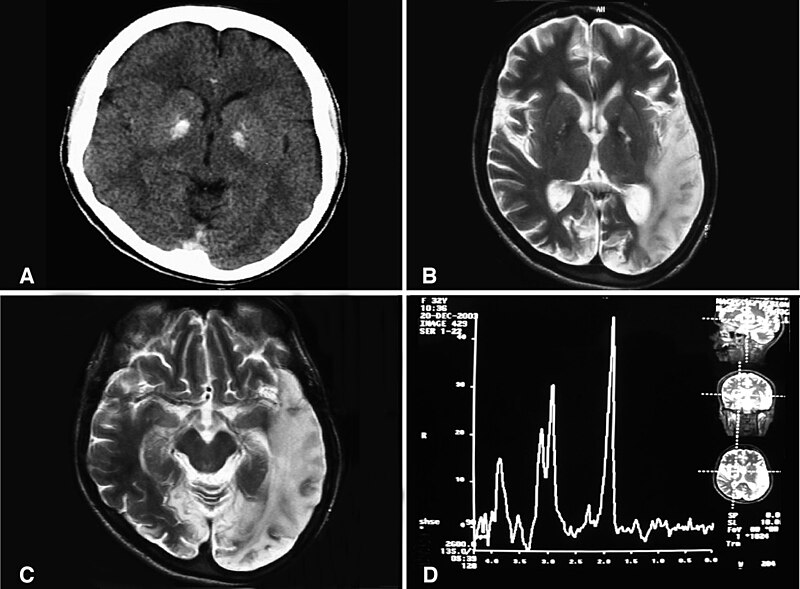
English: (a) A computed tomography brain scan showing bilateral basal ganglia calcification; the cerebellum shows prominent folia indicating mild cerebellar atrophy. (b) Axial T2 brain magnetic resonance image scan showing left temporo-parieto occipital ischemic lesion. (c) Axial T2 brain magnetic resonance image scan showing the extension of the parietal temporal region to the occipital lobe, and also showing a right occipital lesion. (d) Magnetic resonance spectroscopy showing inversion of J-coupling phenomenon at 1.3 ppm, indicating lactate peak. Abu-Amero et al. Journal of Medical Case Reports 2009 3:77 doi:10.1186/1752-1947-3-77 |
| التاريخ | |
| المصدر | A patient with typical clinical features of mitochondrial encephalopathy, lactic acidosis and stroke-like episodes (MELAS) but without an obvious genetic cause: a case report |
| المؤلف | Abu-Amero KK, Al-Dhalaan H, Bohlega S, Hellani A, Taylor RW. |
| الترخيص (إعادة استخدام هذا الملف) |
© 2009 Abu-Amero et al; licensee BioMed Central Ltd. This is an Open Access article distributed under the terms of the Creative Commons Attribution License (https://creativecommons.org/licenses/by/2.0), which permits unrestricted use, distribution, and reproduction in any medium, provided the original work is properly cited. |
هذا الملف مُرخَّص برخصة المشاع الإبداعي العامة المُلزِمة بنسب العمل إلى مُؤَلِّفه 2.0
- يحقُّ لك:
- مشاركة العمل – نسخ العمل وتوزيعه وبثُّه
- إعادة إنتاج العمل – تعديل العمل
- حسب الشروط التالية:
- نسب العمل إلى مُؤَلِّفه – يلزم نسب العمل إلى مُؤَلِّفه بشكل مناسب وتوفير رابط للرخصة وتحديد ما إذا أجريت تغييرات. بالإمكان القيام بذلك بأية طريقة معقولة، ولكن ليس بأية طريقة تشير إلى أن المرخِّص يوافقك على الاستعمال.
تاريخ الملف
اضغط على زمن/تاريخ لرؤية الملف كما بدا في هذا الزمن.
| زمن/تاريخ | صورة مصغرة | الأبعاد | مستخدم | تعليق | |
|---|---|---|---|---|---|
| حالي | 10:20، 28 يناير 2010 | | 1٬200 × 883 (153 كيلوبايت) | CopperKettle | {{Information |Description={{en|1=(a) A computed tomography brain scan showing bilateral basal ganglia calcification; the cerebellum shows prominent folia indicating mild cerebellar atrophy. (b) Axial T2 brain magnetic resonance image scan showing left te |
استخدام الملف
الصفحة التالية تستخدم هذا الملف:
الاستخدام العالمي للملف
الويكيات الأخرى التالية تستخدم هذا الملف:
- الاستخدام في de.wikipedia.org
- الاستخدام في en.wikipedia.org
- الاستخدام في en.wikibooks.org
- الاستخدام في es.wikipedia.org
- الاستخدام في fa.wikipedia.org
- الاستخدام في fr.wikipedia.org
- الاستخدام في it.wikipedia.org
- الاستخدام في la.wikipedia.org
- الاستخدام في ru.wikipedia.org
- الاستخدام في sv.wikipedia.org
- الاستخدام في tr.wikipedia.org
- الاستخدام في www.wikidata.org
- الاستخدام في zh.wikipedia.org