ملف:Hepatocellular carcinoma 1.jpg
المظهر
Hepatocellular_carcinoma_1.jpg (550 × 368 بكسل حجم الملف: 38 كيلوبايت، نوع MIME: image/jpeg)
تاريخ الملف
اضغط على زمن/تاريخ لرؤية الملف كما بدا في هذا الزمن.
| زمن/تاريخ | صورة مصغرة | الأبعاد | مستخدم | تعليق | |
|---|---|---|---|---|---|
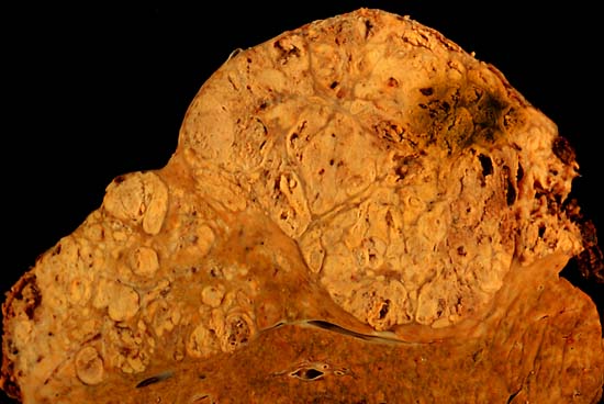
| حالي | 10:14، 5 يونيو 2006 |  | 550 × 368 (38 كيلوبايت) | Patho | {{Information| |Description=Hepatocellular carcinoma This specimen is from a 50ish woman who presented to the hospital with abdominal pain and ascites. The radiologist recovered what appeared to be whole blood on paracentesis. Cytological exam of the blo |
استخدام الملف
ال3 صفحات التالية تستخدم هذا الملف:
الاستخدام العالمي للملف
الويكيات الأخرى التالية تستخدم هذا الملف:
- الاستخدام في ast.wikipedia.org
- الاستخدام في az.wikipedia.org
- الاستخدام في be.wikipedia.org
- الاستخدام في bs.wikipedia.org
- الاستخدام في ca.wikipedia.org
- الاستخدام في cs.wikipedia.org
- الاستخدام في de.wikipedia.org
- الاستخدام في de.wikibooks.org
- الاستخدام في el.wikipedia.org
- الاستخدام في en.wikipedia.org
- Hepatocellular carcinoma
- Alcohol and cancer
- Portal:Medicine/Selected article/50, 2007
- Portal:Medicine/Selected Article Archive (2007)
- Obesity-associated morbidity
- Cirrhosis
- Portal:Viruses/Selected article
- Portal:Viruses/Selected article/10
- User:Daniel Mietchen/Wikidata lists/Items with Disease Ontology IDs
- الاستخدام في eo.wikipedia.org
- الاستخدام في es.wikipedia.org
- الاستخدام في eu.wikipedia.org
- الاستخدام في fa.wikipedia.org
- الاستخدام في fi.wikipedia.org
- الاستخدام في fr.wikipedia.org
- الاستخدام في gl.wikipedia.org
- الاستخدام في he.wikipedia.org
- الاستخدام في hi.wikipedia.org
- الاستخدام في hy.wikipedia.org
- الاستخدام في id.wikipedia.org
- الاستخدام في it.wikipedia.org
- الاستخدام في ja.wikipedia.org
- الاستخدام في kk.wikipedia.org
- الاستخدام في ko.wikipedia.org
اعرض المزيد من الاستخدام العام لهذا الملف.

