مستخدم:Ahmed Aboshama/الانسداد
| الانسداد | |
|---|---|
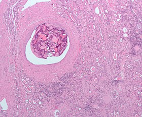
صورة مجهرية لمادة انسداد في الشريان الكلوي. تم استئصال الكلية جراحيا بسبب السرطان. صبغة الهيماتوكسيلين والإيوسين
| |
| تعديل مصدري - تعديل |
الانسداد يشير إلى تمرير وإيداع سدادة في تيار الدم. قد تكون مرضية (وفي هذه الحالة تسمى انسداد وعائي)، مثل الانصمام الرئوي، أو علاجية، كإرقاء لعلاج النزيف أو لعلاج بعض أنواع السرطان عن طريق غلق الأوعية الدموية عمدًا لتجويع خلايا الورم.
في علاج السرطان، بالإضافة لقيام السدادة بإيقاف إمداد الدم للورم، فإنها تحتوي على مكون لمهاجمة الورم كيميائيا أو عن طريق الإشعاع. حين تحمل السدادة دواء علاج كيميائي، تسمى العملية انسداد كيماوي. الانسداد الكيماوي عن طريق قسطرة شريانية هو النموذج المألوف. حين تحمل السدادة دواء مشع تسمى العملية انسداد إشعاعي أو علاج اختياري بالإشعاع الداخلي.
الاستخدامات[عدل]
تتضمن عملية الانسداد السد الاختياري للأوعية الدموية عن طريق إدخال سدادة، أو بكلمات أخرى غلق وعاء دموي بشكل متعمد.
يستخدم الانسداد في علاج قطاع عريض من الحالات المتعلقة بأعضاء مختلفة في جسم الإنسان.
النزف[عدل]
يستخدم العلاج لإغلاق:
- نفث الدم المتكرر
- أم الدم داخل القحف
[1]
- نزف هضمي
- رعاف
- دوالي الخصية
- نزف أولي تال للوضع [2]
- نزف جراحي[3]
- نزف رضحي مثل تمزق الطحال وكسر الحوض
الأورام[عدل]
يستخدم العلاج لإبطاء أو إيقاف إمدادات الدم وبالتالي إنقاص حجم الورم:
- أورام الكلية
- أورام الكبد، بالأخص سرطان الخلية الكبدية. يتم معالجته إما بالإحتشاء الجزئي أو بالانسداد الكيماوي عن طريق القسطرة الشريانية
- الأورام الليفية في الرحم
- تشوه شرياني وريدي
ارتفاع ضغط الدم الخبيث[عدل]
من الممكن أن يكون مفيدا في التعامل مع حالات ارتفاع ضغط الدم الخبيث الناتج عن القصور الكلوي.[4]
أخرى[عدل]
- انسداد الوريد البابي الكبدي قبل استئصال الكبد.[5]
الآلية[عدل]

إن الانسداد عملية جراحية متدنية الإنتهاك. الغرض هو منه سريان الدم إلى منطقة ما في الجسم، مما يمكن بشكل فعال من تصغير حجم ورم أو غلق أم الدم.
يتم تنفيذ هذا الإجراء كإجراء وعائي داخلي عن طريق طبيب أشعة تدخلية متخصص. من الشائع أن يتلقى معظم المرضى العلاج دون أو بالقليل من التخدير، إلا أن ذلك يعتمد بشكل كبير على العضو الذي سيتم منع الدم عنه. يخضع المرضى الذين يجروح جراحة انسداد لغلق الأوعية المخية أو الوريد البابي الكبدي لتخدير كلي عادة.
يتم الوصول للعضو المطلوب عن طريق سلوك التوجيه أو القساطر. وعلى حسب العضو، قد يكون الوصول له صعبًا للغاية ومستهلكًا للوقت. يتم تحديد وضع الشريان أو الوريد السليم الذي يغذي النسيج المرضي عن طريق تصوير الأوعية بالطرح الرقمي. يتم استخدام تلك الصور بعد ذلك كخريطة لطبيب الأشعة التدخلية للوصول إلى الوعاء الدموي الصحيح عن طريق استخدام قسطرة أو سلك مناسبـ بالاعتماد على 'الشكل' التشريحي المحيط.
بمجرد الوصول للمكان، يمكن أن يبدأ العلاج. تكون السدادة الصناعية المستخدمة عادة واحدة مما يلي:
- لفائف: ملف جويليمي القابل للفصل أو الملف المائي
- رقائق
- رغوة
- سدادة
- كريات مجهرية أو خرز
بمجرد إدخال السدادة الصناعية بنجاح، يتم تصوير الأوعية بالطرح الرقمي مرة أخرى للتأكد من نجاح العملية.
المواد المستخدمة[عدل]
مواد الانسداد السائلة - تستخدم في حالات التشوه الشرياني الوريدي، تستطيع هذه المواد السريان من خلال التركيبات الوعائية المعقدة وبالتالي لا يحتاج الجراح أن يوجه قسطرته لكل وعاء دموي. أونيكس هي مثال لمادة انسداد سائلة.
- بوتيل سيانوأكريليت (NBCA) - هذه المادة سائلة دائما وسريعة المفعول، تشبه الصمغ ويتم بيعها المسمى التجاري "الصمغ الخارق" (SuperGlue)، وتتبلمر فور احتكاكها مع الأيونات. كما أنها تتسم بتفاعل منتج للحرارة يقوم بتدمير جدار الوعاء الدموي. نظرا لأن عملية البلمرة تحدث بسرعة كبيرة، فإن تلك المادة تتطلب جراح ماهر. خلال العملية، يجب على الجرح أن يغسل القسطرة قبل وبعد حقن مادة NBCA، وإلا فإنها ستتبلمر داخل القسطرة. كذلك يجب سحب القسطرة سريعًا لكي لا تلتصق بالوعاء الدموي. يمكن مزج الزيت بتلك المادة لإبطاء معدل البلمرة.
- إثيودول - مصنوع من اليود وزيت بذور الخشخاش، وهي مادة لزجة للغاية. تستخدم عادة في عمليات الانسداد الكيماوي، خاصة لسرطان الخلية الكبدية، لكون ذلك الورم يمتص اليود. عمر النصف لتلك المادة هو 5 أيام، ولذلك فهي تسد الأوعية مؤقتًا فقط
Sclerosing agents - These will harden the endothelial lining of vessels. They require more time to react than the liquid embolic agents. Therefore, they cannot be used for large or high-flow vessels.
- ethanol - This permanent agent is very good for treating AVM. The alcohol does need some time to denature proteins of the endothelium and activate the coagulation system to cause a blood clot. Therefore, some surgeons will use a balloon occlusion catheter to stop the blood flow and allow time for ethanol to work. Ethanol is toxic to the system in large quantities and may cause compartment syndrome. In addition, the injections are painful.
- ethanolamine oleate - This permanent agent is used for sclerosing esophageal varices. It contains 2% benzyl alcohol, so it is less painful than ethanol. However it does cause hemolysis and renal failure in large doses.
- sotradecol - This agent is used for superficial lower extremity varicose veins. It has been around for a very long time and is a proven remedy. However, it does cause hyperpigmentation of the region in 30% of patients. It is less painful than ethanol.
Particulate embolic agents - These are only used for precapillary arterioles or small arteries. These are also very good for AVM deep within the body. The disadvantage is that they are not easily targeted in the vessel. None of these are radioopaque, so they are difficult to view with radiologic imaging unless they are soaked in contrast prior to injection.
- Gelfoam hemostasis - Temporarily occludes vessels for five weeks.[6] Works by absorbing liquid and plugging the vessel. Composed of water-insoluble gelatin, so the particles may travel distally and occlude smaller capillaries. One way to localize the injection of gelfoam is to make a gelfoam sandwich. A coil is placed at a precise location, then gelfoam is injected and lodged into the coil.
- polyvinyl alcohol (PVA) - These are permanent agents. They are tiny balls 50-1200 um in size. The particles are not meant to mechanically occlude a vessel. Instead they cause an inflammatory reaction. Unfortunately, they have a tendency to clump together since the balls are not perfectly round. The clump can separate a few days later, failing as an embolic agent.
- Embolization microspheres - These are superior permanent or resorbable particulate embolic agents available in different well-calibrated size ranges for precise occlusion. Embolization microspheres may comprise additional functionality such as drug loading and elution capability, specific mechanical properties, imageability or radioactivity
Mechanical occlusion devices - These fit in all vessels. They also have the advantage of accuracy of location; they are deployed exactly where the catheter ends.
- coils - These are used for AVF, aneurysms, or trauma. They are very good for fast-flowing vessels because they immediately clot the vessel. They are made from platinum or stainless steel. They induce clots due to the Dacron wool tails around the wire. The coil itself will not cause mechanical occlusion. Since it is made of metal, it is easily seen in radiographic images. The disadvantage is that large coils can disrupt the radiographic image. The coil may also lose its shape if the catheter is kinked. Also, there is a small risk of dislodging from the deployed location.
- detachable balloon - Treats AVF and aneurysms. These balloons are simply implanted in a target vessel, then filled with saline through a one-way valve. The blood stops and endothelium grows around the balloon until the vessel fibroses. The balloon may be hypertonic relative to blood and hence rupture and fail, or it may be hypotonic and shrink, migrating to a new location.
المميزات[عدل]
- جراحة متدنية الإنتهاك
- لا تندب
- خطر ضئيل للعدوى
- لا استخدام أو استخدام نادر للتخدير
- زمن تعافي أسرع
- نسبة نجاح عالية مقارنة بالأساليب الأخرى
- يحافظ على الخصوبة والسلامة التشريحية
العيوب[عدل]
- تعتمد نسبة النجاح على المستخدم
- خطورة وصول السدادات إلى أنسجة سليمة مسببة قرحة معدية أو قرحة الاثنا عشر.[7] توجد طرق وآليات وأجهزة تقلل من حدوث هذا النوع من الآثار الجانبية.
- ليست مناسبة للجميع
- التكرار أكثر احتمالا
انظر أيضا[عدل]
مراجع[عدل]
- ^ Jiang B، Paff M، Colby GP، Coon AL، Lin LM (سبتمبر 2016). "Cerebral aneurysm treatment: modern neurovascular techniques". Stroke Vasc Neurol. ج. 1 ع. 3: 93–100. DOI:10.1136/svn-2016-000027. PMC:5435202. PMID:28959469.
- ^ Chauleur C، Fanget C، Tourne G، Levy R، Larchez C، Seffert P (يوليو 2008). "Serious primary post-partum hemorrhage, arterial embolization and future fertility: a retrospective study of 46 cases". Hum. Reprod. ج. 23 ع. 7: 1553–9. DOI:10.1093/humrep/den122. PMID:18460450.
- ^ Whittingham-Jones، P.؛ Baloch، I.؛ Miles، J.؛ Ferris، B (2010). "Persistent haemarthrosis following total knee arthroplasty caused by unrecognised arterial injury". Grand Rounds. ج. 10: 51–54. DOI:10.1102/1470-5206.2010.0010 (غير نشط 15 يناير 2017). مؤرشف من الأصل في 2010-10-24.
{{استشهاد بدورية محكمة}}: الوسيط غير المعروف|deadurl=تم تجاهله (مساعدة) ويحتوي الاستشهاد على وسيط غير معروف وفارغ:|month=(مساعدة)صيانة الاستشهاد: وصلة دوي غير نشطة منذ 2017 (link) - ^ Alhamid N، Alterky H، Othman MI (يناير 2013). "Renal artery embolization for managing uncontrolled hypertension in a kidney transplant candidate". Avicenna J Med. ج. 3 ع. 1: 23–5. DOI:10.4103/2231-0770.112791. PMC:3752858. PMID:23984264.
{{استشهاد بدورية محكمة}}: صيانة الاستشهاد: دوي مجاني غير معلم (link) - ^ Madoff DC، Hicks ME، Vauthey JN، Charnsangavej C، Morello FA، Ahrar K، Wallace MJ، Gupta S (2002). "Transhepatic portal vein embolization: anatomy, indications, and technical considerations". Radiographics. ج. 22 ع. 5: 1063–76. DOI:10.1148/radiographics.22.5.g02se161063. PMID:12235336.
- ^ Gelfoam at Pfizer
- ^ Carretero C، Munoz-Navas M، Betes M، Angos R، Subtil JC، Fernandez-Urien I، De la Riva S، Sola J، Bilbao JI، de Luis E، Sangro B (يونيو 2007). "Gastroduodenal injury after radioembolization of hepatic tumors". The American Journal of Gastroenterology. ج. 102 ع. 6: 1216–20. DOI:10.1111/j.1572-0241.2007.01172.x. PMID:17355414.
روابط خارجية[عدل]
[[تصنيف:علم الأشعة التدخلي]] [[تصنيف:جراحة]]

